

QTc Interval and QT Dispersion in Patients with Thalassemia Major: Electrocardiographic (EKG) and Echocardiographic Evaluation
- PMID: 20567638
- PMCID: PMC2884339
- DOI: 10.4137/cmc.s4472
QTc Interval and QT Dispersion in Patients with Thalassemia Major: Electrocardiographic (EKG) and Echocardiographic Evaluation
Abstract
Background: Doppler echocardiographic studies in patients with beta-Thalassemia Major (beta-TM) had shown different patterns of left ventricle (LV) systolic and diastolic dysfunctions.
Aim: This cross-sectional study was designed to study the LV systolic and diastolic function in patients with beta-TM using Pulsed Doppler (PD) Echocardiogram and assess the QTc interval and QT dispersion (QTd) on 12 leads ECG.
Method: All patients were evaluated clinically as well as by echocardiography and 12 leads ECG. The study included patients with beta-TM (n = 38, age 15.7 +/- 8.9 years), compared with an age-matched healthy control group (n = 38, age 15.9 +/- 8.9 years).
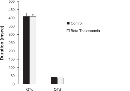
Results: In 38 patients with beta-TM Compared with healthy control group, The QTc interval and the QTd dispersion on ECG were increased with no significant difference mode echo showed that beta-TM patients have thicker LV septal wall index (0.659 +/- 0.23 vs. 0.446 +/- 0.219 cm/M(2), P < 0.001), posterior wall index (0.659 +/- 0.235 vs. 0.437 +/- 0.214 cm/M(2), P < 0.01), and larger LVEDD index is (3.99 +/- 0.48 vs. 2.170 +/- 0.57 cm/M(2). P < 0.05). Pulsed Doppler showed high LV trans-mitral E wave velocity index (70.818 +/- 10.139 vs. 57.532 +/- 10.139, P < 0.05) and E/A ratio (1.54 vs.1.23, P < 0.01). The duration of deceleration time index (DT) and isovolumic relaxation time index (IVRT) were significantly shorter in patients with beta-TM (150.234 +/- 20.0.23 vs. 167.123 +/- 167.123 +/- 19.143 msec/M(2), P < 0.01) and (60.647 +/- 6.77 vs. 75.474 +/- 5.83 msec/M(2), P < 0.001), respectively. The tricuspid valve velocity in patients with beta-TM was significantly higher than controls (2.993 +/- 0.569 vs. 1.93 +/- 0.471 m/sec, respectively, P < 0.01), with calculated pulmonary artery pressure of 2.4 times the control (36.0 vs. 14.8 mmHg). However, the LVEF% or fractional shortening were not significantly different.
Conclusion: In this study, beta-thalassemia major patients compared with controls have differences of QT dispersion and corrected QT interval that is of no statistical significance. A significantly thicker LV wall and LV diastolic filling indices are suggestive of restrictive diastolic pattern. These data indicate that LV diastolic abnormalities compromised initially in patients with beta-thalassemia major.
Keywords: Bahrain; QT dispersion; beta-thalassemia major; pulsed echo Doppler.
Figures
References
-
- Fujita S. Congenital hemolytic anemia–hemoglobin abnormality–thalassemia. Nippon Rinsho. 1996;54:2454–9. - PubMed
-
- Fosburg MT, Nathan DG. Treatment of Cooley’s anemia. Blood. 1990;76:435–44. - PubMed
-
- 3-Ehlers KH, Levin AR, Markenson AL, Marcus JR, Klein AA, Hilgartner MW, et al. Longitudinal study of cardiac function in thalassemia major. Ann N Y Acad Sci. 1980;344:397–404. - PubMed
-
- 4-Hahalis G, Manolis AS, Apostolopoulos D, Alexopoulos D, Vagenakis AG, Zoumbos NC. Right ventricular cardiomyopathy in beta-thalassaemia major. Eur Heart J. 2002;23:147–56. - PubMed
-
- Al-Arrayed S, Hafadh N, Amin S, Al-Mukhareq H, Sanad H. Student screening for inherited blood disorders in Bahrain. Eastern Mediterranean Health Journal. 2003;9(3):344–52. - PubMed
LinkOut - more resources
Full Text Sources
