

Management of cutaneous tumors with mohs micrographic surgery
- PMID: 20567701
- PMCID: PMC2884874
- DOI: 10.1055/s-0028-1095884
Management of cutaneous tumors with mohs micrographic surgery
Abstract
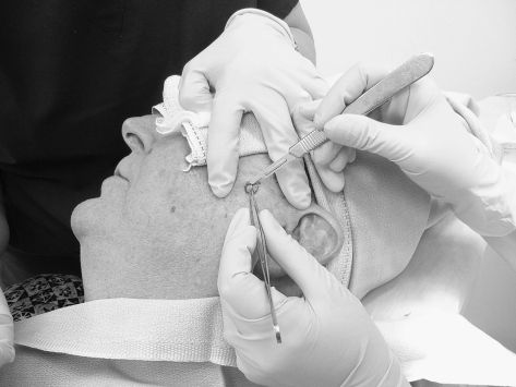
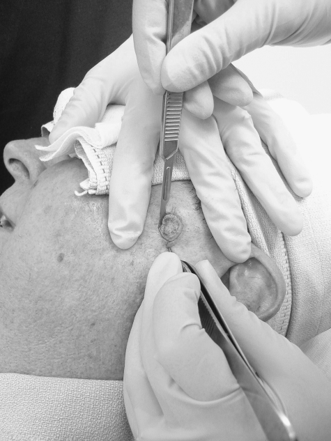
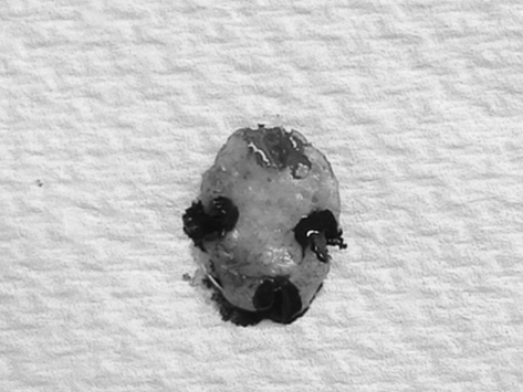
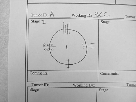
Since the inception of Mohs micrographic surgery in the 1930s, this technique has proved its utility in the treatment of cutaneous tumors. This review describes the technique of Mohs micrographic surgery and the various indications for which it is used. We discuss the use of Mohs micrographic surgery for the following cutaneous tumors: basal cell carcinoma, squamous cell carcinoma, melanoma in situ, dermatofibrosarcoma protuberans, Merkel cell carcinoma, microcystic adnexal carcinoma, atypical fibroxanthoma, and sebaceous carcinoma. Mohs micrographic surgery is cost effective in the U.S. health care system because billing for the surgeon-pathologist and laboratory processing is bundled together. However, Mohs micrographic surgery may be more expensive in European systems because the Mohs technique surgeon, pathologist, and laboratory fees may be billed separately.
Keywords: Mohs micrographic surgery; cutaneous oncology; skin cancer.
Figures
Similar articles
-
Mohs micrographic surgery: a review of indications, technique, outcomes, and considerations.An Bras Dermatol. 2021 May-Jun;96(3):263-277. doi: 10.1016/j.abd.2020.10.004. Epub 2021 Mar 24. An Bras Dermatol. 2021. PMID: 33849752 Free PMC article. Review.
-
Mohs micrographic surgery in rare cutaneous tumors: a retrospective study at a Brazilian tertiary university hospital.An Bras Dermatol. 2023 Jan-Feb;98(1):36-46. doi: 10.1016/j.abd.2022.01.009. Epub 2022 Nov 8. An Bras Dermatol. 2023. PMID: 36369200 Free PMC article.
-
Mohs micrographic surgery: a treatment method for many non-melanocytic skin cancers.Dermatol Online J. 2020 Apr 15;26(4):13030/qt8zr4f9n4. Dermatol Online J. 2020. PMID: 32621679
-
Mohs micrographic surgery: established uses and emerging trends.Oncology (Williston Park). 2005 Aug;19(9):1165-71; discussion 1171-2, 1175. Oncology (Williston Park). 2005. PMID: 16255133
-
Current progress of immunostains in Mohs micrographic surgery: a review.Dermatol Surg. 2008 Dec;34(12):1621-36. doi: 10.1111/j.1524-4725.2008.34339.x. Epub 2008 Oct 13. Dermatol Surg. 2008. PMID: 19018832 Review.
Cited by
-
An Analysis of Lidocaine Usage in the Treatment of Squamous Cell Carcinoma.Cureus. 2023 Feb 28;15(2):e35614. doi: 10.7759/cureus.35614. eCollection 2023 Feb. Cureus. 2023. PMID: 37021063 Free PMC article. Review.
-
Management of dermatofibrosarcoma protuberans of the face using lower trapezius musculocutaneous pedicle flap reconstruction: a case report.J Surg Case Rep. 2018 Jun 15;2018(6):rjy089. doi: 10.1093/jscr/rjy089. eCollection 2018 Jun. J Surg Case Rep. 2018. PMID: 29942467 Free PMC article.
-
Mohs micrographic surgery: a review of indications, technique, outcomes, and considerations.An Bras Dermatol. 2021 May-Jun;96(3):263-277. doi: 10.1016/j.abd.2020.10.004. Epub 2021 Mar 24. An Bras Dermatol. 2021. PMID: 33849752 Free PMC article. Review.
-
A practical update of surgical management of merkel cell carcinoma of the skin.ISRN Surg. 2013;2013:850797. doi: 10.1155/2013/850797. Epub 2013 Jan 30. ISRN Surg. 2013. PMID: 23431473 Free PMC article.
-
Dermatofibrosarcoma protuberans: a case report and review of the literature.Hippokratia. 2016 Jan-Mar;20(1):80-83. Hippokratia. 2016. PMID: 27895450 Free PMC article.
References
-
- Mohs F E. Mohs micrographic surgery: a historical perspective. Dermatol Clin. 1989;7:609–611. - PubMed
-
- Brodland D G, Amonette R, Hanke C W, Robins P. The history and evolution of Mohs micrographic surgery. Dermatol Surg. 2000;26:303–307. - PubMed
-
- Mohs F E. Chemosurgery: a microscopically controlled method of cancer excision. Arch Surg. 1941;42:279.
-
- Mohs F E. In: Roenigk RK, Roenigk HH, editor. Dermatologic Surgery: Principles and Practice. New York, NY: Marcel Dekker; 1989. History of Mohs micrographic surgery. pp. 783–789.
-
- Tromovitch T A, Stegman S J. Microscopically controlled excision of skin tumors: chemosurgery (Mohs): fresh tissue technique. Arch Dermatol. 1974;110:231–232. - PubMed
