. 2008 Nov;22(4):269-80.
doi: 10.1055/s-0028-1095886.

Lip reconstruction

Affiliations

Lip reconstruction

Donald Baumann et al. Semin Plast Surg. 2008 Nov.

Abstract

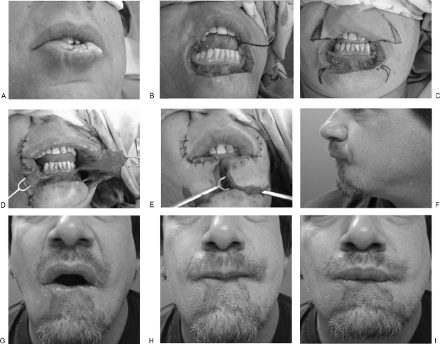
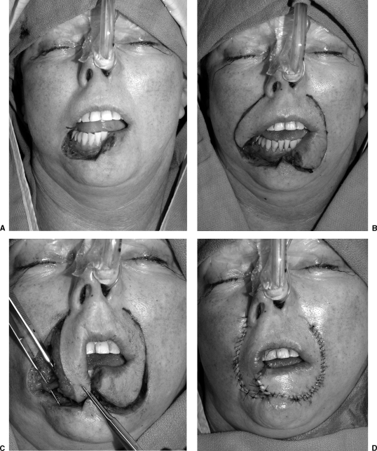
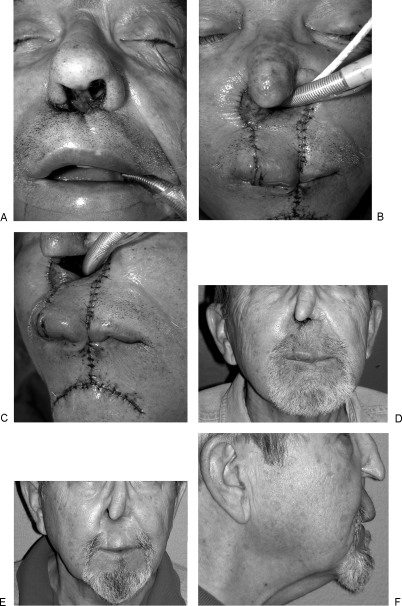
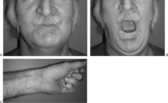
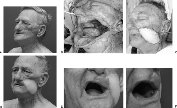
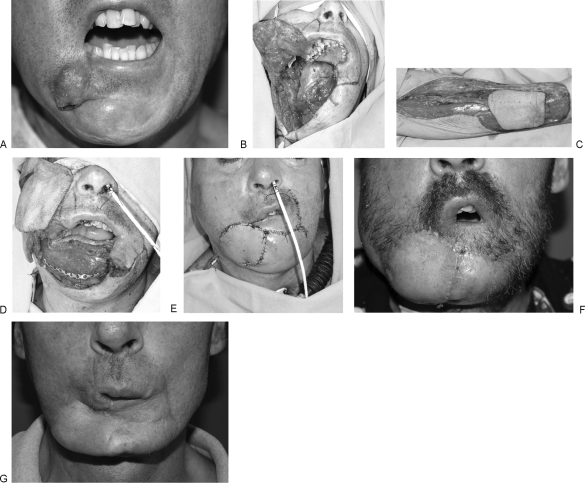
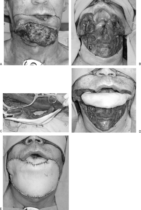
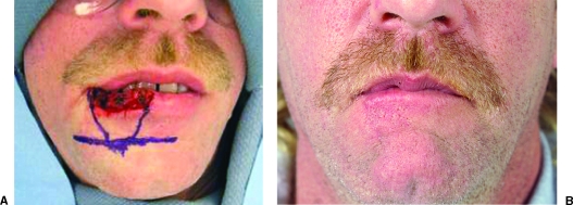
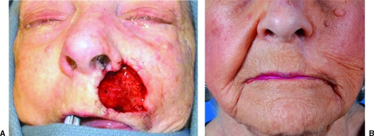
Lip reconstruction poses a particular challenge to the plastic surgeon in that the lips are the dynamic center of the lower third of the face. Their role in aesthetic balance, facial expression, speech, and deglutination is not replicated by any other tissue substitute. The goals of lip reconstruction are both functional and aesthetic, and the surgical techniques employed are often overlapping. This discussion will focus on lip defects with significant tissue loss that require flap reconstruction. Flaps described include Webster-Bernard cheek advancement flaps, Abbe cross-lip flaps, Karapandzic rotation advancement flaps, and single and dual free-flap lip reconstructions. The principles and techniques described are broadly applicable to other flap designs that are required to meet both the aesthetic and functional goals of lip reconstruction.

Keywords: Abbe flap; Karapandzic flap; Lip reconstruction; Webster-Bernard flap; free flap.


Figures

Figure 1
Figure 1
Squamous cell carcinoma of the lower lip reconstructed with the Webster-Bernard flap. (A) Preoperative view. (B) Total lower lip defect; note the commissures are preserved. (C) Planned markings for bilateral cheek advancement with the Webster-Bernard flap. (D) Flaps mobilized; note the extent of mucosa recruited for creation of the vermilion. (E) Flap inset. (F–I) Twelve-month follow-up.
Figure 2
Figure 2
T2 N0 squamous cell carcinoma subtotal lower lip defect reconstructed with a Karapandzic flap. (A) Extirpative defect. Right commissure marked. (B) Flap design along nasolabial fold. (C) Flaps mobilized. Neurovascular pedicle isolated and preserved. (D) Flap inset. (All photos copyright © 2007, Matthew Hanasono, M.D.)
Figure 3
Figure 3
Recurrent squamous cell carcinoma of nasal vestibule and upper lip treated previously with radiation. (A) View of planned resection of nasal vestibule, septum, and upper lip. (B) Abbe flap with addition of mental skin to resurface defect of nasal floor. (C) Width of flap design was based on wider tissue requirement of nasal floor than of upper lip defect. (D) Result at 2 weeks before flap division. (E) Three-month follow up. (F) Lateral view; projection of the central lip preserved. (All photos copyright © 2007, Roman Skoracki, M.D.)
Figure 4
Figure 4
T4 N0 squamous cell carcinoma of nasal cavity, columella, and upper lip. (A) Extirpative defect after total rhinectomy, palatectomy, bilateral maxillectomies, and resection of upper lip resulting in large confluent oro-nasal cavity. (B) Palate and upper lip reconstructed with folded radial forearm flap. The flap was oriented such that the radial and ulnar borders were inset posteriorly allowing the pedicle to travel laterally to the left facial vessels. A palmaris sling anchored to the zygomatic bone supported the flap. (All photos copyright © 2007, Donald Baumann, M.D.)
Figure 5
Figure 5
Eight-month follow-up after radial forearm free-flap reconstruction of lower lip defect. (A) Lip in repose. Flap was supported with fascia lata sling anchored to the modiolus. Of note, the palmaris tendon was absent. (B) Lip circumference symmetrically preserved. (C) Healed donor site. (All photos copyright © 2007, Roman Skoracki, M.D.)
Figure 6
Figure 6
Recurrent squamous cell carcinoma after previous reconstruction with vertical rectus abdominus myocutaneous flap and radiation therapy. (A) Preoperative appearance. Note the tethered scar retraction and lateral displacement of the commissure. Intraoral soft tissues are also contracted and fixed. (B) Full-thickness cheek defect including the lateral element of the upper and lower lips and commissure. (C) Reconstruction with dual paddled anterolateral thigh flap and fascia lata sling anchored to the zygomatic bone. The upper and lower lip elements were advanced to create a new commissure supported by the underlying ALT flap. (D) Follow-up at 8 weeks. (E) Improved mouth opening. (F) Reconstructed commissure maintains anatomic position upon mouth opening. (All photos copyright © 2007, Donald Baumann, M.D.)
Figure 7
Figure 7
T4 N0 squamous cell carcinoma of the mandible eroding through to external skin and undermining the lower lip. (A) Preoperative appearance. (B) Defect after hemimandibulectomy and resection of lower lip and chin-cheek skin. The defect required intraoral bone and soft tissue reconstruction that was provided by a fibula osteocutaneous flap. A radial forearm flap provided external skin coverage. (C) Radial forearm skin island. (D) Fibula inset and radial forearm flap revascularized. Lip reconstruction was planned with a unilateral Karapandzic flap. (E) Intraoperative result after flap inset. (F) Result at 4 weeks. Reconstructed right lower lip is well supported by the soft tissue framework below; however, the shortened lip length has led to reduction in oral opening. (G) Result at 8 months after completion of radiation therapy. Note the improved contour of radial forearm flap. (All photos copyright © 2007, Matthew Hanasono, M.D.)
Figure 8
Figure 8
Exophytic squamous cell carcinoma of mandible and lower third of the face. (A) Preoperative appearance. (B) Extirpative defect including angle-to-angle mandibulectomy, resection of floor of mouth, lower lip, chin and neck skin. This defect required a dual free-flap reconstruction, bone and soft tissue intraoral reconstruction, and a large soft tissue flap for external neck and chin coverage. Given the extent of lip and intraoral resection, there were limited options for the lip reconstruction. The fibula skin island was used for resurfacing of the lower lip in addition to intraoral coverage. (C) Harvest of fibula with large skin paddle. (D) Fibula inset with soft tissue platform for lip reconstruction. (E) Intraoperative result with ALT flap resurfacing the chin and cheek and neck skin. (All photos copyright © 2007, Matthew Hanasono, M.D.)
Figure 1
Figure 1
Interoperative/postoperative views of a nearly 50% lower lip defect closed by wedge excision/closure.
Figure 2
Figure 2
30% skin only upper lip defect reconstructed with a peri-alar crescentric advancement flap.

References

    1. Adams W, Beran S. Lip, cheek, and scalp reconstruction; hair replacement. Selected Readings in Plastic Surgery. 2001;15:2–16.
    1. Kroll S. In: Kroll S, editor. Reconstructive Plastic Surgery for Cancer. St. Louis, MO: Mosby Year Book; 1996. Lip reconstruction. pp. 201–209.
    1. Webster R C, Coffey R J, Kelleher R E. Total and partial reconstruction of the lower lip with innervated muscle-bearing flaps. Plast Reconstr Surg Transplant Bull. 1960;25:360–371. - PubMed
    1. Closmann J J, Pogrel M A, Schmidt B L. Reconstruction of perioral defects following resection for oral squamous cell carcinoma. J Oral Maxillofac Surg. 2006;64:367–374. - PubMed
    1. Langstein H N, Robb G L. Lip and perioral reconstruction. Clin Plast Surg. 2005;32:431–445. - PubMed