Vascularized growth plate transfer for distal radius reconstruction
- PMID: 20567713
- PMCID: PMC2884881
- DOI: 10.1055/s-2008-1081402
Vascularized growth plate transfer for distal radius reconstruction
Abstract
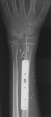
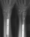
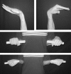
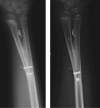
Distal radius reconstruction in children should meet two requests: restoration of some joint function and preservation of the physiologic growth of the segment. None of the conventional options is likely to successfully achieve both goals. Conversely, a vascularized transfer of the proximal fibula including the growth plate provides enough bone stock for diaphyseal reconstruction, an articular surface for joint function, and the potential for longitudinal growth. From 1992 to 2006, eight children ranging in age between 2 and 10 years underwent a vascularized transfer of the proximal fibula for distal radius reconstruction after bone sarcoma resection. The follow-up ranges were between 1 year and 15 years. All the grafts were harvested based on the anterior tibial artery. Seven cases with a follow-up longer than 2 years have been evaluated both clinically and radiographically. All the grafts survived and had a satisfactory growth after the transplant. The functional outcome has been satisfactory, and the range of motion of the reconstructed wrist has been nearly normal in all cases but one. Proximal fibular epiphyseal transfer was an effective procedure for distal radius reconstruction in children who underwent tumor resection. Refinements in the operative technique have increased the reliability of this reconstructive option, which might be safely used also in congenital and posttraumatic disorders.
Keywords: Epiphyseal transfer; bone reconstruction; distal radius reconstruction; pediatric microsurgery.
Figures


Similar articles
-
Vascularized proximal fibular epiphyseal transfer for distal radial reconstruction.J Bone Joint Surg Am. 2005 Sep;87 Suppl 1(Pt 2):237-46. doi: 10.2106/JBJS.E.00295. J Bone Joint Surg Am. 2005. PMID: 16140797
-
Long Term Results of Epiphyseal Transplant in Distal Radius Reconstruction in Children.Handchir Mikrochir Plast Chir. 2015 Apr;47(2):83-9. doi: 10.1055/s-0035-1547304. Epub 2015 Apr 21. Handchir Mikrochir Plast Chir. 2015. PMID: 25897577 Review.
-
Vascularized proximal fibular epiphyseal transfer for distal radial reconstruction.J Bone Joint Surg Am. 2004 Jul;86(7):1504-11. doi: 10.2106/00004623-200407000-00021. J Bone Joint Surg Am. 2004. PMID: 15252100
-
Clinical and radiological results of vascularized fibular epiphyseal transfer after bone tumor resection in children.Orthop Traumatol Surg Res. 2020 Nov;106(7):1319-1324. doi: 10.1016/j.otsr.2020.03.037. Epub 2020 Oct 10. Orthop Traumatol Surg Res. 2020. PMID: 33051168
-
Vascularized proximal fibular epiphyseal transfer: two cases.Orthop Traumatol Surg Res. 2012 Oct;98(6):728-32. doi: 10.1016/j.otsr.2012.05.009. Epub 2012 Sep 19. Orthop Traumatol Surg Res. 2012. PMID: 23000036
Cited by
-
New options for vascularized bone reconstruction in the upper extremity.Semin Plast Surg. 2015 Feb;29(1):20-9. doi: 10.1055/s-0035-1544167. Semin Plast Surg. 2015. PMID: 25685100 Free PMC article.
-
Vascularized Fibular Epiphyseal Transfer for Pediatric Limb Salvage: Review of Applications and Outcomes.Plast Reconstr Surg Glob Open. 2023 Oct 18;11(10):e5354. doi: 10.1097/GOX.0000000000005354. eCollection 2023 Oct. Plast Reconstr Surg Glob Open. 2023. PMID: 37859637 Free PMC article.
-
The vascularized fibular graft in the pediatric upper extremity: a durable, biological solution to large oncologic defects.Sarcoma. 2013;2013:321201. doi: 10.1155/2013/321201. Epub 2013 Oct 7. Sarcoma. 2013. PMID: 24222724 Free PMC article.
-
Complication of osteo reconstruction by utilizing free vascularized fibular bone graft.BMC Surg. 2020 Oct 2;20(1):216. doi: 10.1186/s12893-020-00875-9. BMC Surg. 2020. PMID: 33008361 Free PMC article. Review.
-
Reconstruction of the Distal Radius following Tumour Resection Using an Osteoarticular Allograft.Sarcoma. 2013;2013:318767. doi: 10.1155/2013/318767. Epub 2013 Apr 3. Sarcoma. 2013. PMID: 23690732 Free PMC article.
References
-
- Innocenti M, Ceruso M, Manfrini M, et al. Free vascularized growth-plate transfer after bone tumor resection in children. J Reconstr Microsurg. 1998;14:137–143. - PubMed
-
- Innocenti M, Delcroix L, Romano G F. Epiphyseal transplant: harvesting technique of the proximal fibula based on the anterior tibial artery. Microsurgery. 2005;25:284–292. - PubMed
-
- Innocenti M, Delcroix L, Manfrini M, et al. Vascularized proximal fibular epiphyseal transfer for distal radial reconstruction. J Bone Joint Surg Am. 2005;87(Suppl 1 Pt 2):237–246. - PubMed
-
- Wenger H L. Transplantation of epiphyseal cartilage. Arch Surg. 1954;50:148–151.
-
- Wilson J N. Epiphyseal transplantation. A clinical study. J Bone Joint Surg Am. 1966;48:245–256. - PubMed

