Vascularized bone grafts and their applications in the treatment of carpal pathology
- PMID: 20567715
- PMCID: PMC2884887
- DOI: 10.1055/s-2008-1081404
Vascularized bone grafts and their applications in the treatment of carpal pathology
Abstract
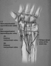
Vascularized bone grafts (VBGs) are techniques in the management of certain types of carpal pathology. VBGs have traditionally been advocated for conditions including delayed and nonunion of fractures and avascular necrosis. The most common indications for VBG have been for scaphoid nonunion, lunatomalacia (Kienböck's disease), and osteonecrosis of the scaphoid (Preiser's disease). Advantages over NVBG have been established. VBGs provide improved blood flow, osteocyte preservation, and accelerated healing rates. Local pedicled VBGs are the most commonly used methods. They are technically less demanding than are free VBGs and are associated with less morbidity. Commonly used donor grafts arise from the dorsal vasculature of the wrist and include the 1,2 intercompartmental supraretinacular artery (1,2 ICSRA), the 2,3 ICSRA, the fourth extensor compartment artery (fourth ECA), and the fifth ECA. A 4 + 5 ECA combination graft has been described to provide a longer pedicle. In managing osteonecrosis, most surgeons would agree that VBG should be reserved for carpal bones with an intact cartilaginous shell and no collapse. In treating scaphoid pathology, indications for VBG include fractures/nonunions with proximal pole avascular necrosis and/or small proximal pole fragments.
Keywords: Vascular bone grafting; nonunion; osteonecrosis.
Figures


Similar articles
-
Vascularized bone grafts for the treatment of carpal bone pathology.Hand (N Y). 2013 Mar;8(1):27-40. doi: 10.1007/s11552-012-9479-0. Hand (N Y). 2013. PMID: 24426890 Free PMC article.
-
[Pedicled vascularized bone grafts from the dorsum of the distal radius for treatment of scaphoid nonunions].Oper Orthop Traumatol. 2009 Nov;21(4-5):373-85. doi: 10.1007/s00064-009-1908-z. Oper Orthop Traumatol. 2009. PMID: 20058117 Clinical Trial. German.
-
Bone Grafting for Scaphoid Nonunions: Is Free Vascularized Bone Grafting Superior for Scaphoid Nonunion?Hand (N Y). 2019 Mar;14(2):217-222. doi: 10.1177/1558944717736397. Epub 2017 Oct 27. Hand (N Y). 2019. PMID: 29078719 Free PMC article.
-
The Efficacy of Vascularized Bone Grafts in the Treatment of Scaphoid Nonunions and Kienbock Disease: A Systematic Review in 917 Patients.J Hand Microsurg. 2019 Apr;11(1):6-13. doi: 10.1055/s-0038-1677318. Epub 2018 Dec 26. J Hand Microsurg. 2019. PMID: 30911206 Free PMC article. Review.
-
The Use of the Proximal Hamate as an Autograft for Proximal Pole Scaphoid Fractures: Clinical Outcomes and Biomechanical Implications.Hand Clin. 2019 Aug;35(3):287-294. doi: 10.1016/j.hcl.2019.03.007. Epub 2019 May 11. Hand Clin. 2019. PMID: 31178087 Review.
Cited by
-
Reconstruction of Large Skeletal Defects: Current Clinical Therapeutic Strategies and Future Directions Using 3D Printing.Front Bioeng Biotechnol. 2020 Feb 12;8:61. doi: 10.3389/fbioe.2020.00061. eCollection 2020. Front Bioeng Biotechnol. 2020. PMID: 32117940 Free PMC article. Review.
-
Treatment of AVN-Induced Proximal Pole Scaphoid Nonunion Using a Fifth and Fourth Extensor Compartmental Artery as a Vascularized Pedicle Bone Graft: A Retrospective Case Series.Med Sci Monit. 2024 May 19;30:e944553. doi: 10.12659/MSM.944553. Med Sci Monit. 2024. PMID: 38762751 Free PMC article.
-
Mandatory Surgeon Skills for Care of the Mutilated Hand.J Hand Surg Glob Online. 2024 Aug 22;7(2):314-318. doi: 10.1016/j.jhsg.2024.07.007. eCollection 2025 Mar. J Hand Surg Glob Online. 2024. PMID: 40182865 Free PMC article. Review.
-
Vascularized bone grafts for the treatment of carpal bone pathology.Hand (N Y). 2013 Mar;8(1):27-40. doi: 10.1007/s11552-012-9479-0. Hand (N Y). 2013. PMID: 24426890 Free PMC article.
-
Prognostic factors in the treatment of carpal scaphoid non-unions.Eur J Orthop Surg Traumatol. 2017 Jan;27(1):3-9. doi: 10.1007/s00590-016-1886-4. Epub 2016 Nov 28. Eur J Orthop Surg Traumatol. 2017. PMID: 27896458 Review.
References
-
- Jupiter J B, Gerhard H D, Guerrero J, et al. Treatment of segmental defects of the radius with use of the vascularized osteoseptocutaneous fibular autogenous graft. J Bone Joint Surg Am. 1997;79:542–550. - PubMed
-
- Weiland A J, Kleinert H E, Kutz J E, et al. Free vascularized bone grafts in surgery of the upper extremity. J Hand Surg [Am] 1979;4:129–144. - PubMed
-
- Gabl M, Lutz M, Reinhart C, et al. Stage 3 Kienböck's disease: reconstruction of the fractured lunate using a free vascularized iliac bone graft and external fixation. J Hand Surg [Br] 2002;27:369–373. - PubMed
-
- Gabl M, Reinhart C, Lutz M, et al. Vascularized bone graft from the iliac crest for the treatment of nonunion of the proximal part of the scaphoid with an avascular fragment. J Bone Joint Surg Am. 1999;81:1414–1428. - PubMed
-
- Doi K, Oda T, Soo-Heong T, et al. Free vascularized bone graft for nonunion of the scaphoid. J Hand Surg [Am] 2000;25:507–519. - PubMed

