

Reconstruction of osteomyelitis defects
- PMID: 20567733
- PMCID: PMC2884901
- DOI: 10.1055/s-0029-1214163
Reconstruction of osteomyelitis defects
Abstract
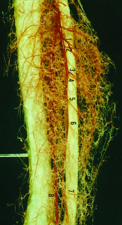
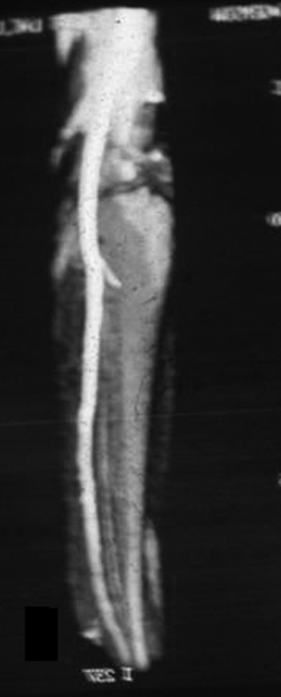
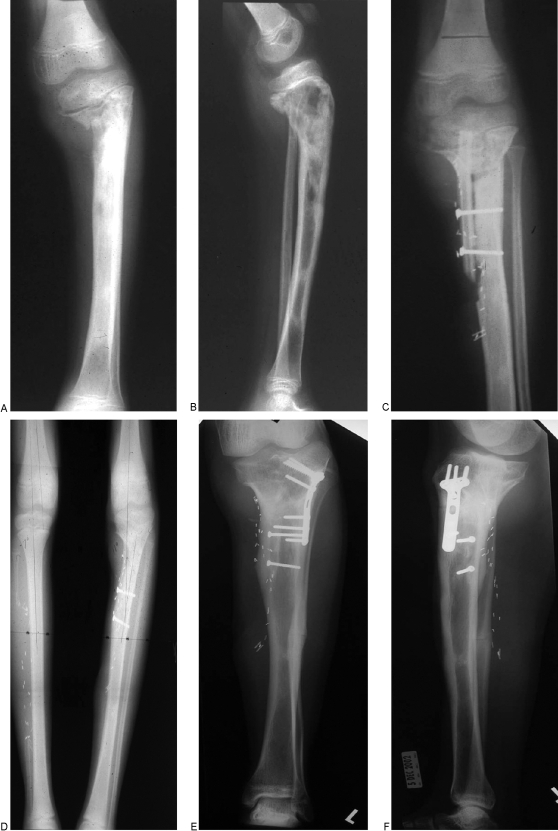
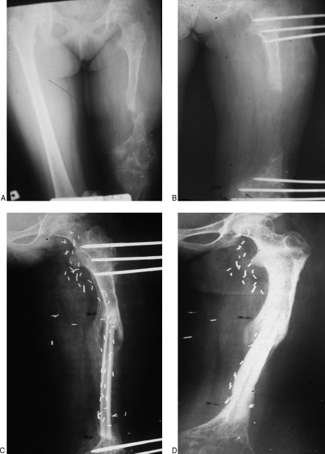
Reconstruction of large skeletal defects secondary to osteomyelitis remains a challenging problem. Osteomyelitis can result from a variety of etiologies; most often, it is a consequence of trauma to a long bone. Despite advances in antibiotic therapy, treatment of chronic osteomyelitis requires adequate surgical debridement, which can often lead to large soft tissue and bone loss. Free vascularized bone can be used to reconstruct large skeletal defects greater than 6 cm or bone defects of smaller size that failed to heal with nonvascularized bone grafting. The length, cortical strength, and anatomic configuration of the free vascular fibular graft make it an ideal bone graft to bridge extremity defects, and it can be transferred with skin, fascia, and muscle to fill soft tissue defects in the recipient site.
Keywords: Osteomyelitis; osteocutaneous flaps; skeletal defects; vascularized fibular graft.
Figures

Similar articles
-
Vascularized bone grafts for post-traumatic defects in the upper extremity.Arch Plast Surg. 2021 Jan;48(1):84-90. doi: 10.5999/aps.2020.00969. Epub 2021 Jan 15. Arch Plast Surg. 2021. PMID: 33503750 Free PMC article.
-
Reconstruction of large skeletal defects due to osteomyelitis with the vascularized fibular graft in children.J Bone Joint Surg Am. 2007 Oct;89(10):2233-40. doi: 10.2106/JBJS.E.01319. J Bone Joint Surg Am. 2007. PMID: 17908901
-
One-stage treatment and reconstruction of Gustilo Type III open tibial shaft fractures with a vascularized fibular osteoseptocutaneous flap graft.J Orthop Trauma. 2010 Dec;24(12):745-51. doi: 10.1097/BOT.0b013e3181d88a07. J Orthop Trauma. 2010. PMID: 21063216
-
Role of vascularized bone grafts in lower extremity osteomyelitis.Orthop Clin North Am. 2007 Jan;38(1):37-49, vi. doi: 10.1016/j.ocl.2006.10.005. Orthop Clin North Am. 2007. PMID: 17145293 Review.
-
Free vascularized fibular grafting in combination with a locking plate for the reconstruction of a large tibial defect secondary to osteomyelitis in a child: a case report and literature review.J Pediatr Orthop B. 2010 Jan;19(1):66-70. doi: 10.1097/BPB.0b013e328331c340. J Pediatr Orthop B. 2010. PMID: 19898254 Review.
Cited by
-
Bony Hypertrophy in Vascularized Fibular Grafts.Hand (N Y). 2022 Jan;17(1):106-113. doi: 10.1177/1558944719895784. Epub 2020 Jan 27. Hand (N Y). 2022. PMID: 31984803 Free PMC article.
-
Surgical treatment options for septic non-union of the tibia: two staged operation, Flow-through anastomosis of FVFG, and continuous local intraarterial infusion of heparin.Fukushima J Med Sci. 2016 Dec 16;62(2):83-89. doi: 10.5387/fms.2016-5. Epub 2016 Jul 30. Fukushima J Med Sci. 2016. PMID: 27477992 Free PMC article.
-
Acute on Chronic Osteomyelitis of the Humerus - A Case Report on a Neglected Case of Chronic Osteomyelitis with Bone Cement In Situ.J Orthop Case Rep. 2022;12(5):40-44. doi: 10.13107/jocr.2022.v12.i05.2808. J Orthop Case Rep. 2022. PMID: 36660155 Free PMC article.
-
Mycobacterium fortuitum osteomyelitis of the cuboid bone treated with CERAMENT G and V: a case report.J Bone Jt Infect. 2022 Jul 25;7(4):163-167. doi: 10.5194/jbji-7-163-2022. eCollection 2022. J Bone Jt Infect. 2022. PMID: 36032800 Free PMC article.
-
Both Bone Forearm Infected Nonunion: Report of a One-Bone Free Fibula Flap Salvage and Literature Review.Hand (N Y). 2020 Jul;15(4):NP51-NP56. doi: 10.1177/1558944719857168. Epub 2019 Jun 19. Hand (N Y). 2020. PMID: 31215792 Free PMC article. Review.
References
-
- Lew D P, Waldvogel F A. Osteomyelitis. N Engl J Med. 1997;336:999–1007. - PubMed
-
- Moore J R, Weiland A J. Vascularized tissue transfer in the treatment of osteomyelitis. Clin Plast Surg. 1986;13:657–662. - PubMed
-
- Patzakis M J, Zalavras C G. Chronic posttraumatic osteomyelitis and infected nonunion of the tibia: current management concepts. J Am Acad Orthop Surg. 2005;13:417–427. - PubMed
-
- Tu Y K, Yen C Y, Yeh W L, Wang I C, Wang K C, Ueng W N. Reconstruction of posttraumatic long bone defect with free vascularized bone graft: good outcome in 48 patients with 6 years' follow-up. Acta Orthop Scand. 2001;72:359–364. - PubMed
-
- Manolagas S C. Birth and death of bone cells: basic regulatory mechanisms and implications for the pathogenesis and treatment of osteoporosis. Endocr Rev. 2000;21:115–137. - PubMed
LinkOut - more resources
Full Text Sources
Other Literature Sources
