. 2009 May;23(2):119-31.
doi: 10.1055/s-0029-1214164.

Reconstruction of osteomyelitis defects of the craniofacial skeleton

Affiliations

Reconstruction of osteomyelitis defects of the craniofacial skeleton

Gary E Decesare et al. Semin Plast Surg. 2009 May.

Abstract

Osteomyelitis of the craniofacial skeleton closely resembles osteomyelitis elsewhere in the body in its pathophysiology and medical management; subsequent reconstruction after debridement remains distinctly challenging. The goals of reconstruction must include the restoration of the complex and readily visible morphology of the cranium and face, as well as the adequate return of vital sensory, expressive, and digestive functions. In this article, the various reconstructive modalities will be discussed including pedicled and nonpedicled flaps with or without an osseous component, nonvascularized bone grafts, alloplastic implants, and bone regeneration using protein therapy. Although reconstruction of craniofacial defects after osteomyelitis commonly proves formidable, the satisfactory return of form and function remains a plausible reconstructive goal.

Keywords: Osteomyelitis; craniofacial; head and neck; mandible; midface; reconstruction; skull.


Figures
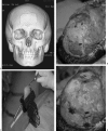
Figure 1
Figure 1
(A) CT scan of frontal bone defect from Pott's puffy tumor, temporarily repaired with titanium mesh. (B) Frontal bone defect (top of image); harvested full-thickness calvarial graft from parietal bone (bottom of image). (C) Full-thickness calvarial graft in process of being split with saw. (D) Outer table graft repairing frontal defect (top of image); inner table graft replaced orthotopically (bottom of image).
Figure 2
Figure 2
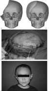
(A) Near hemi-calvarial defect in young child. (B) Custom-made porous polyethylene implant. (C) Calvarial implant rigidly fixed in place. (D) Postoperative result.
Figure 3
Figure 3
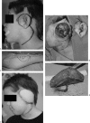
(A) Left orbital/skull base defect. (B) Temporalis muscle flap raised. (C) Temporalis muscle flap mobilized into skull base defect. (D) Left orbital/skull base defect filled with temporalis muscle flap.
Figure 4
Figure 4
(A) Planned resection of temporal skull base lesion. (B) Temporal skull base defect. (C) Anterolateral thigh flap donor site marked. (D) Anterolateral thigh free flap. (E) Postoperative temporal skull defect with anterolateral thigh free flap reconstruction.
Figure 5
Figure 5
(A) Mucormycosis infection of the facial soft tissues and maxillary sinus. (B) Staged resection of facial and maxillary sinus mucormycosis. The patient expired secondary to systemic disease prior to reconstruction.
Figure 6
Figure 6
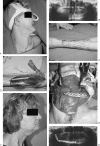
(A) Preoperative right-sided mandibular defect. (B) Panorex of right-sided mandibular defect. (C) Markings for radial forearm osteocutaneous free flap. (D) Elevation of radial forearm osteocutaneous free flap. (E) Intraoperative inset of radial forearm osteocutaneous free flap and reconstruction plate. (F) Postoperative result: lateral view. (G) Postoperative Panorex.
Figure 7
Figure 7
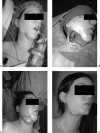
(A) Patient after dental infection, debridement, and segmental mandibular defect initially treated with reconstruction plate now exposed. (B) Intraoperative surgical defect. (C) Intraoperative inset: skin paddle of the fibular free flap used for cutaneous reconstruction. (D) Six-month follow-up: postoperative new after partial skin paddle removal.

Similar articles

Cited by

References

    1. Nelaton A. Elements De Pathologie Chirurgicale. Paris, France: Germer-Bailliere; pp. 1844–1859.
    1. David D J, Cooter R D. Craniofacial infection in 10 years of transcranial surgery. Plast Reconstr Surg. 1987;80:213–225. - PubMed
    1. Fearon J A, Yu J, Bartlett S P, Munro I R, Chir B, Whitaker L. Infections in craniofacial surgery: a combined report of 567 procedures from two centers. Plast Reconstr Surg. 1997;100:862–868. - PubMed
    1. Barker F G., II Efficacy of prophylactic antibiotics for craniotomy: a meta-analysis. Neurosurgery. 1994;35:484–490. discussion 491–482. - PubMed
    1. Verbon A, Husni R N, Gordon S M, Lavertu P, Keys T F. Pott's puffy tumor due to Haemophilus influenzae: case report and review. Clin Infect Dis. 1996;23:1305–1307. - PubMed