. 2010 May;6(5):888-93.
doi: 10.1039/b917876g. Epub 2010 Feb 9.

High sensitivity detection of cancer in vivo using a dual-controlled activation fluorescent imaging probe based on H-dimer formation and pH activation

Affiliations

High sensitivity detection of cancer in vivo using a dual-controlled activation fluorescent imaging probe based on H-dimer formation and pH activation

Mikako Ogawa et al. Mol Biosyst. 2010 May.

Abstract

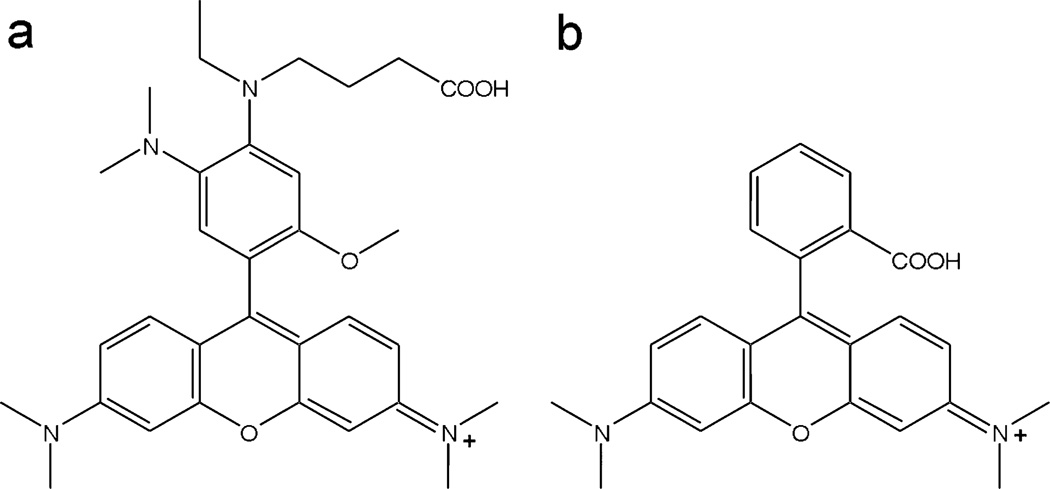
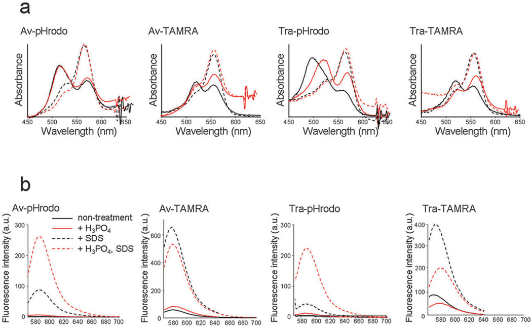
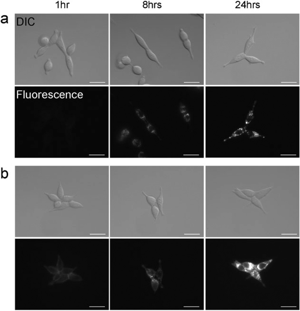
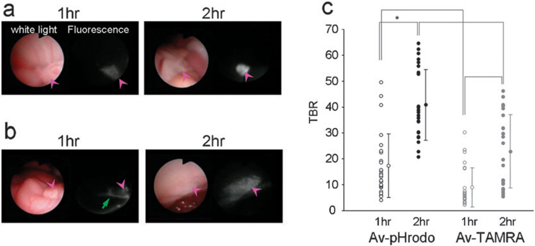
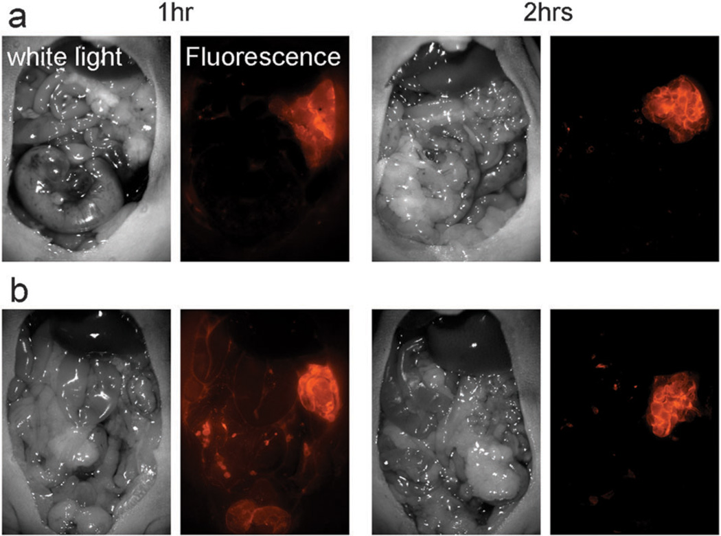
The key to improving the sensitivity of in vivo molecular imaging is to increase the target-to-background signal ratio (TBR). Optical imaging has a distinct advantage over other molecular imaging methods in that the fluorescent signal can be activated at the target thus reducing background signal. Previously, we found that H-dimer formation quenches fluorescence of xanthene fluorophores, and among these, TAMRA had the highest quenching ratio. Another approach to lowering background signal is to employ pH activation based on the photon-induced electron transfer (PeT) theory. We hypothesized that combining these two strategies could lead to greater quenching capacity than was possible with either probe alone. A pH-sensitive fluorophore, pHrodo or TAMRA was conjugated to the cancer targeting molecules, avidin (Av) and trastuzumab (Tra). As expected, both pHrodo and TAMRA formed H-dimers when conjugated to avidin or antibody and the dimerization resulted in efficient fluorescence quenching. In addition, pHrodo conjugated probes showed pH-dependent fluorescence activation. When the probes were used in an in vivo animal model, fluorescence endoscopy with Av-pHrodo depicted tumors with high TBR 1 h and 2 h after injection. Av-TAMRA also visualized tumors 1 h and 2 h after the injection, however, TBR was lower due to the background signal from non-specific binding 1 h after the injection as well as background fluorescence from the unbound agent. Thus, we demonstrate that a dual-controlled activatable optical probe based on the combination of H-dimer formation and pH activation can achieve high TBR at early time points during in vivo molecular imaging.


Figures

Fig. 1
Fig. 1
The proposed structure of pHrodo (a) and the structure of TAMRA (b).
Fig. 2
Fig. 2
(a) Absorbance spectrum of each conjugate without SDS (solid line) and with SDS (dashed line), and at neutral pH (black) and acidic (red) conditions. The blue shifted peak without SDS represents H-dimer formation of fluorophores. Both pHrodo and TAMRA showed H-dimer formation. (b) Fluorescence spectra of each conjugate without SDS (solid line) and with SDS (dashed line). The fluorescence was increased by SDS addition (H-dimer dissociation) in both pHrodo and TAMRA conjugates. In addition, pHrodo probes showed fluorescence activation by lowering pH, and this effect resulted in the large fluorescence activation capacity for pHrodo conjugates.
Fig. 3
Fig. 3
Fluorescence microscopy as well as differential interference contrast (DIC) images of Tra-pHrodo (a) or Tra-TAMRA (b) in 3T3/HER2+ tumor cells. The cell-surface fluorescence signal was not detected with Tra-pHrodo, but a weak signal was detected with Tra-TAMRA. Fluorescent dots were detected within the cytoplasm 8 h after incubation at 37 °C with both conjugates. Bars = 20 µm.
Fig. 4
Fig. 4
In vivo fluorescence endoscopic images in peritoneal tumor bearing mice with Av-pHrodo (a) or Av-TAMRA (b). The pink arrow heads show the tumor nodules. The tumors were visualized with both Av-pHrodo and Av-TAMRA 1 h after the injection, but fluorescence from excess injectate in the peritoneal cavity (green arrow) was also detected. Two hours after the imaging probe injection, the tumor nodules were clearly visualized by both Av-pHrodo and Av-TAMRA, although the background signal was lower for Av-pHrodo. The measured tumor-to-background ratios (TBRs) are summarized in (c). The highest TBR was accomplished by Av-pHrodo 2 h after the injection. (*p < 0.001, Mann-Whitney U test).
Fig. 5
Fig. 5
Fluorescence spectral images of the peritoneal cavity for Av-pHrodo (a) and Av-TAMRA (b). The results were consistent with endoscopic images. Tumors were detected with a low background signal for Av-pHrodo, but the injectate fluorescence was detected with Av-TAMRA at 1 h after the injection.

Similar articles

Cited by

References

    1. Weissleder R, Pittet MJ. Nature. 2008;452:580–589. - PMC - PubMed
    1. Pierce MC, Javier DJ, Richards-Kortum R. Int. J. Cancer. 2008;123:1979–1990. - PMC - PubMed
    1. Kosaka N, Ogawa M, Sato N, Choyke PL, Kobayashi H. J. Invest. Dermatol. 2009;129:2818–2822. - PMC - PubMed
    1. Kobayashi H, Ogawa M, Kosaka N, Choyke PL, Urano Y. Nanomedicine. 2009;4:411–419. - PMC - PubMed
    1. Longmire M, Kosaka N, Ogawa M, Choyke PL, Kobayashi H. Cancer Sci. 2009;100:1099–1104. - PMC - PubMed

Publication types