Review
. 2010 Nov;19(11):1824-36.
doi: 10.1007/s00586-010-1476-9. Epub 2010 Jun 22.

Sagittal plane deformity: an overview of interpretation and management

Affiliations
Review

Sagittal plane deformity: an overview of interpretation and management

Pierre Roussouly et al. Eur Spine J. 2010 Nov.

Abstract

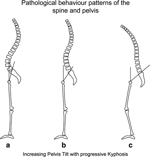
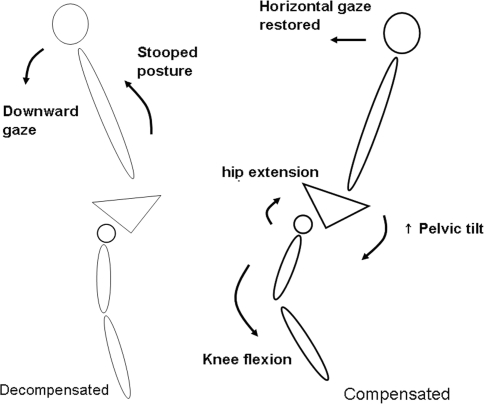
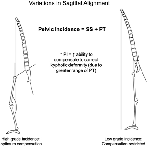
The impact of sagittal plane alignment on the treatment of spinal disorders is of critical importance. A failure to recognise malalignment in this plane can have significant consequences for the patient not only in terms of pain and deformity, but also social interaction due to deficient forward gaze. A good understanding of the principles of sagittal balance is vital to achieve optimum outcomes when treating spinal disorders. Even when addressing problems in the coronal plane, an awareness of sagittal balance is necessary to avoid future complications. The normal spine has lordotic curves in the cephalad and caudal regions with a kyphotic curve in between. Overall, there is a positive correlation between thoracic kyphosis and lumbar lordosis. There are variations on the degree of normal curvature but nevertheless this shape allows equal distribution of forces across the spinal column. It is the disruption of this equilibrium by pathological processes or, as in most cases, ageing that results in deformity. This leads to adaptive changes in the pelvis and lower limbs. The effects of limb alignment on spinal posture are well documented. We now also know that changes in pelvic posture also affect spinal alignment. Sagittal malalignment presents as an exaggeration or deficiency of normal lordosis or kyphosis. Most cases seen in clinical practise are due to kyphotic deformity secondary to inflammatory, degenerative or post-traumatic disorders. They may also be secondary to infection or tumours. There is usually pain and functional disability along with concerns about self-image and social interaction due to inability to maintain a horizontal gaze. The resultant pelvic and lower limb posture is an attempt to restore normal alignment. Addressing this complex problem requires detailed expertise and awareness of the potential pitfalls surrounding its treatment.


Figures

Fig. 1
Fig. 1
a Chin-brow vertical angle, b Kyphosis tilt angle, c C7 plumb line (sagittal vertical axis), d Horizontal distance (HD) of displaced sagittal vertical axis from reference point on sacral end plate
Fig. 2
Fig. 2
The 3 main pelvic parameters. When measuring pelvic incidence, the reference point is the centre of the femoral head (if the femoral heads superimpose) or the mid point of the line connecting the femoral heads (if they do not superimpose)
Fig. 3
Fig. 3
a Spino-pelvic angle and b Spino-sacral angle
Fig. 4
Fig. 4
Variations of spinal alignment. Types I–IV. Note the thoracolumbar split (as a measure of length ratio of the thoracic and lumbar curves) in each type: a Type I 80:20, b type II 60:40 c type III 50:50, d 20:80
Fig. 5
Fig. 5
Pathological behaviour pattern of pelvis in presence of kyphosis shows a normal posture, b abnormal posture, and c compensatory posture. Pelvic tilt increases in order to maintain an upright posture
Fig. 6
Fig. 6
Sagittal plane imbalance. Decompensated posture on the left versus compensated posture on the right. Note change in lower limb profile with extension of hip joints and flexion of knees
Fig. 7
Fig. 7
Demonstration of relationship between pelvic incidence and pelvic tilt in compensatory adjustment for kyphosis. Smaller pelvic incidence indicates less compensatory ability
Fig. 8
Fig. 8
Polysegmental wedge osteotomies showing resection of the lamina and facets. This leaves segmental gaps posteriorly at each level which are closed with compression to shorten the posterior column and correct deformity
Fig. 9
Fig. 9
Pedicle subtraction osteotomy. a Lateral view of vertebra with wedge removed, b posterior vertebral wall with posterior arch and pedicles removed, c posterior view of vertebra after wedge excision. Re-alignment is effected by apposition of the remaining upper and lower portions of the vertebra by compression
Fig. 10
Fig. 10
Change in lordotic profile of lumbar spine with shift in apex when osteotomy performed at different levels (L3 vs. L4). Note the shorter lumbar curve when the osteotomy is performed at L4
Fig. 11
Fig. 11
Restoration of sagittal alignment is measured by the distance between the plumb lines (A) when the osteotomy is performed at B which is further away and distal to the apex of the deformity therefore requiring a smaller angle of correction C
Fig. 12
Fig. 12
Restoration of sagittal alignment with osteotomy (X) closer to apex of deformity. Angle of correction C1 required to give same amount of improvement in sagittal correction (A) is greater. In both cases, the plumb line (sagittal vertical axis, SVA1) shifts posteriorly (SVA) with restoration of alignment
Fig. 13
Fig. 13
Posterior vertebral column resection diagram to demonstrate margins of resection: 1 Posterior arch, 2 Adjacent lamina, 3 Transverse processes/ribs, 4 Discs, 5 Vertebral body. The anterior longitudinal ligament is left intact to act as a restraint to overcorrection
Fig. 14
Fig. 14
Smith Petersen osteotomy: Posterior osteotomies (black lines) with deformity correction through disc space and anterior longitudinal ligament. The correction hinges on the middle and posterior columns. There is a risk of great vessel rupture with this method

References

    1. Berthonnaud E, Dimner J, Roussouly P, Labelle H. Analysis of the sagittal balance of the spine and pelvis using shape and orientation parameters. J Spine Disord. 2004;18(1):40–47. doi: 10.1097/01.bsd.0000117542.88865.77. - DOI - PubMed
    1. Berven SH, Deviren V, Smith JA, Emami A, Hu SS, Bradford DS. Management of fixed sagittal plane deformity: results of the transpedicular wedge resection osteotomy. Spine. 2001;26(18):2036–2043. doi: 10.1097/00007632-200109150-00020. - DOI - PubMed
    1. Boachie-Adjei O, Bradford DS. Vertebral column resection and arthrodesis for complex spinal deformities. J Spinal Disord. 1991;4(2):193–202. doi: 10.1097/00002517-199106000-00010. - DOI - PubMed
    1. Boesker EH, Moe JH, Winter RB, Koop SE. A determination of the normal thoracic kyphosis: a roentgenographic study of 121 normal children. J Pediatr Orthop. 2000;20:796–798. - PubMed
    1. Bradford DS, Schumacher WL, Lonstein JE. Ankylosing spondylitis: experience in surgical management of 21 patients. Spine. 1987;12:238–243. doi: 10.1097/00007632-198704000-00008. - DOI - PubMed