. 2010 Dec;16(4):639-67.
doi: 10.1007/s11948-010-9201-y. Epub 2010 Jun 22.

Avoiding twisted pixels: ethical guidelines for the appropriate use and manipulation of scientific digital images

Affiliations

Avoiding twisted pixels: ethical guidelines for the appropriate use and manipulation of scientific digital images

Douglas W Cromey. Sci Eng Ethics. 2010 Dec.

Abstract

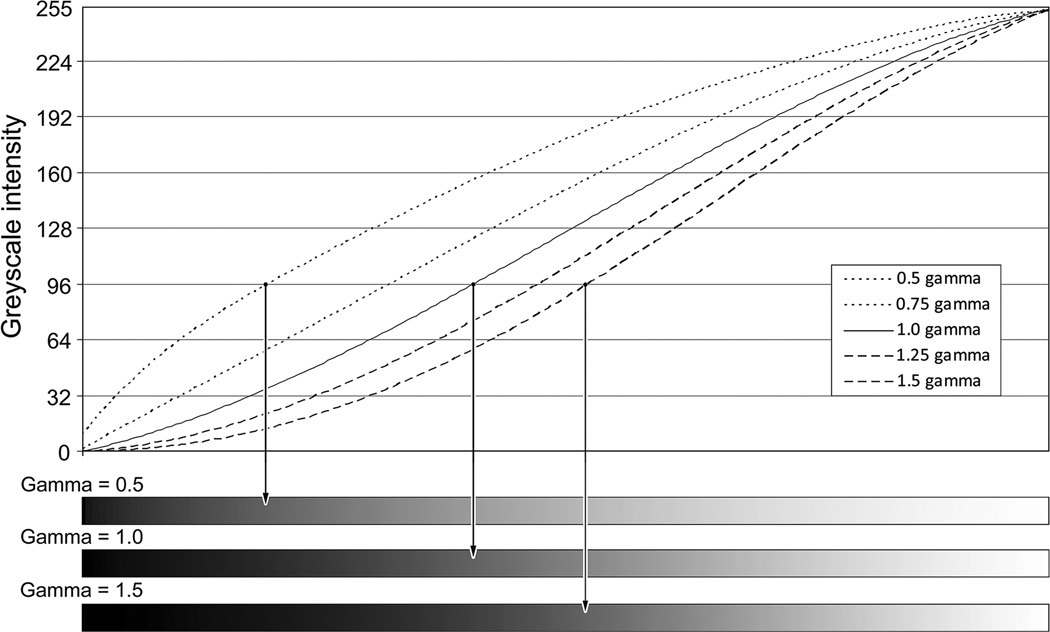
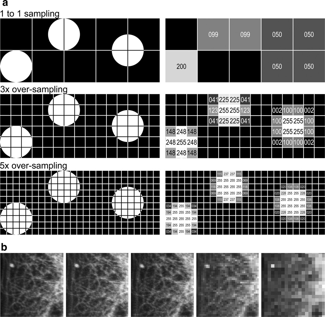
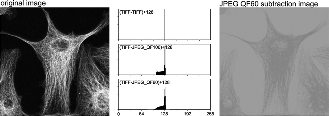
Digital imaging has provided scientists with new opportunities to acquire and manipulate data using techniques that were difficult or impossible to employ in the past. Because digital images are easier to manipulate than film images, new problems have emerged. One growing concern in the scientific community is that digital images are not being handled with sufficient care. The problem is twofold: (1) the very small, yet troubling, number of intentional falsifications that have been identified, and (2) the more common unintentional, inappropriate manipulation of images for publication. Journals and professional societies have begun to address the issue with specific digital imaging guidelines. Unfortunately, the guidelines provided often do not come with instructions to explain their importance. Thus they deal with what should or should not be done, but not the associated 'why' that is required for understanding the rules. This article proposes 12 guidelines for scientific digital image manipulation and discusses the technical reasons behind these guidelines. These guidelines can be incorporated into lab meetings and graduate student training in order to provoke discussion and begin to bring an end to the culture of "data beautification".


Figures

Fig. 1
Fig. 1
Histograms and images. Confocal microscopy image of a mollusk embryo at the 4-cell stage, showing the cytoskeleton of a single cell. The image is courtesy of James Cooley and Lisa Nagy, University of Arizona. Unprocessed image—The original 8-bit (256 shades of grey) image. No post-acquisition image processing was performed. The intensity of this image ranges from the darkest pixel value of 11 to the brightest pixel value of 186. The intensity histogram scale, by convention, runs from darkest on the left, to brightest on the right. There are no true black or white pixels in this image. Appropriately processed image—The same image, after an appropriate contrast/histogram stretch. Using the Photoshop levels tool, the value of 11 from the original image was re-mapped to black (=0) and the value of 186 was remapped to white (=255). Note that the shape of the histogram is essentially the same as in the unprocessed image. The gaps in the histogram are a result of the contrast/histogram stretch. This is generally considered an acceptable image processing step. With color images that will be used for illustrative purposes, it can be useful to apply the levels tool in this way to each of the red, green, and blue channels. If the color images are for quantitative use, or if the relationships of the intensities or colors to one another will be interpreted in any way, this is not recommended. Over-processed image—The same image, this time with a contrast/histogram stretch that was too aggressive. Using the Photoshop levels tool, the value of 20 from the original image was re-mapped to black and the value of 145 was re-mapped to white. Compare the shape of the intensity histogram with the original. Note that the data at each end of the histogram have changed. The data at the ends of the original histogram have been truncated, creating the spikes at black and white (arrows). Nothing scientific can be inferred about these white and black pixels, as their relationship to the rest of the data has been lost. This is a common image processing mistake, arrived at by a number of different techniques, as users try to create striking, “contrasty” images. Boxes—50 × 50 pixel areas from the same area in the un-processed and overprocessed images above. The areas have been enlarged using the Photoshop CS2 nearest neighbor algorithm. Box 1—Note the loss of information in the darkest pixels. The loss is easier to see in the intensity histogram than in the image (arrow). Scientists are often not interested in this end of the histogram; however, backgrounds that are too “clean” do not accurately represent real biology. Box 2—Note the over-saturation of many of the brightest pixels in this image (arrow). Since many journals are using on-line images as the “journal of record”, the data of record are missing some of the fine detail that may be of more interest to the reader than they were to the authors
Fig. 2
Fig. 2
Gamma. A greyscale gradient from pure black to white was created using Adobe Photoshop CS3 with an assumed gamma level of 1.0. Gamma values of 1.5, 1.25, 0.75 and 0.5 were applied to the gradient using ImageJ 1.37 (Rasband 1997–2009). A line profile analysis was performed at each gamma level using ImageJ. The curves were plotted and smoothed with a polynomial trendline in MS Excel and the trendlines are presented in the graph. The x axis is the distance along the gradient and the y axis is the 8-bit greyscale intensity value. To determine how much a greyscale intensity value has been shifted by the application of gamma (a non-linear function), find the place where a y axis gridline intersects the gamma = 1 trendline (solid line), and follow it over to the left or right along the gridline to one of the other gamma trendlines. Gamma enhances the mid-range intensity values more than the extremes of dark or light. Below the graph are examples of the gradients with gamma values = 0.5, 1.0 and 1.5. The arrows show where the greyscale intensity of 96 falls on each gradient
Fig. 3
Fig. 3
a Sampling—theoretical. This illustration shows three bright (intensity = 255) spots that are aligned in different ways with the pixels on a sensor. Each spot is the same size; in each row the detectors are sampling the spot at higher frequencies. The left-most spot can be accurately measured at all three sampling levels. The other two spots give widely varying measurements that are more accurate as the sampling level increases. At 10× oversampling (not shown), all three spots measure exactly 10 pixels in both the x and y axes. In this example, it is assumed that there is no space between the pixels, though in reality this is never the case. In places where the spot did not fill the pixel, the mean intensity was measured using ImageJ (Rasband 1997–2009). This is similar to how a CCD camera treats a partially-illuminated pixel. These reduced-intensity pixels demonstrate how a feature is displayed using aliasing. If a user is too aggressive when processing an image, the aliased pixels can become bright enough to look like a real feature. This would be particularly problematic in the 1 to 1 sampling image, since the three spots could be blended together by over-processing, thus leading to a possible misinterpretation of the data. Note, this example assumes maximum brightness in every portion of the spot. In reduced light conditions (e.g., fluorescence microscopy), these assumptions may not be correct. b Sampling—example. A small area from the same field of view as that used in Fig. 4 was imaged on a Zeiss LSM 510 confocal microscope at 2048 × 2048 pixels, 1024 × 1024, 512 × 512, 256 × 256 and 128 × 128 pixels using the same optical magnification. The areas have been enlarged using the Photoshop CS3 nearest neighbor algorithm and each image was individually contrast stretched and a gamma adjustment of 1.1 was applied to clearly show the pixilation
Fig. 4
Fig. 4
JPEG compression. The left image is of an Invitrogen/Molecular Probes (Eugene, OR) FluoCells #2 slide stained for anti-a-tubulin, captured with a Zeiss LSM 510 confocal microscope. The file was exported from the native Zeiss LSM file format to TIFF and cropped slightly. The TIFF image was then saved as a JPEG file in Photoshop CS3 (save for web and devices) using either the 100 quality factor (minimal compression) or the 60 quality factor (higher compression, with a barely noticeable level of artifacts). Since digital images are a representation of the numerical intensity values for each pixel, image comparison can be performed using simple mathematics. If two images are identical, subtracting one image from the other should yield a product that equals 0. Since JPEG images can change individual pixel values above or below their original value, subtracting two images and then adding 128 will prevent negative values from being missed. A value of 128 (mid-grey) indicates that there has been no change from the original image to the JPEG image. The top intensity histogram is the result when the TIFF image was subtracted from itself and 128 was added. Since the images were identical, the resulting image has a histogram where all the values are an intensity of 128. The middle intensity histogram is of the image that results from subtracting the JPEG (QF 100) from the TIFF image and adding 128. The bottom intensity histogram was made in a similar manner using the JPEG (QF 60) image. Where these two histograms deviate from 128, the original intensity data has been compromised. The image on the right is the image that was created when the JPEG (QF 60) was subtracted from the original TIFF image and then 128 was added to the result. This image was not processed additionally. There is a considerable amount of information that was altered in the JPEG (QF60) image
Fig. 5
Fig. 5
Moiré. The top image is of a portion of a diatom acquired using a Zeiss LSM 510 confocal microscope in differential interference contrast mode. The bottom image is a demonstration image of the kind of moiré artifact that can occur in images that have repeating structures and have been incorrectly down-sampled or were initially undersampled. Note the curved artifacts (arrows) in the diatom in the bottom image. This artifact is the result of aliasing the periodic features in the diatom. This is a somewhat extreme example; however, users need to be aware that down-sampling an image (i.e., reducing the total number of pixels in X and Y) can reduce the information content of an image, and may introduce unwanted, and unnoticed, artifacts. Noise was removed from the top image using Photoshop CS2’s despeckle filter and a conservative contrast stretch performed to enhance the image. The bottom image is a screen capture taken when the above image was viewed at 33% on screen and then the captured image was enlarged using the Photoshop CS3 nearest neighbor algorithm

Comment in

Similar articles

Cited by

References

    1. Abbott A. Forged images lead to German inquiry. Nature. 1997;387(6632):442. - PubMed
    1. Abraham E. Update on the AJRCCM-2007. American Journal of Respiratory and Critical Care Medicine. 2007;175(3):207–208. - PubMed
    1. Abraham E, Adler KB, Shapiro SD, Leff AR. The ATS journals’ policy on image manipulation. Proceedings of the American Thoracic Society. 2008;5(9):869. - PubMed
    1. Adler J. Veracity of raw images can also come into question. Nature. 2005;435(7043):736. - PubMed
    1. Adobe Systems. Adobe Photoshop 7.0, lesson 17-setting up your monitor for color management. San Jose, CA: Adobe Systems Inc.; 2002.

Publication types

LinkOut - more resources