Review
. 2010 Aug;239(8):2141-8.
doi: 10.1002/dvdy.22350.

Toward an understanding of the role of notochordal cells in the adult intervertebral disc: from discord to accord

Affiliations
Review

Toward an understanding of the role of notochordal cells in the adult intervertebral disc: from discord to accord

Makarand V Risbud et al. Dev Dyn. 2010 Aug.

Abstract

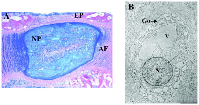
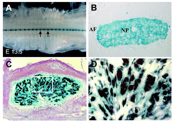
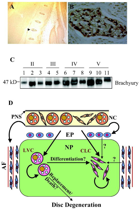
The goal of this mini-review is to address the long standing argument that the pathogenesis of disc disease is due to the loss and/or the replacement of the notochordal cells by other cell types. We contend that, although cells of different size and morphology exist, there is no strong evidence to support the view that the nucleus pulposus contains cells of distinct lineages. Based on lineage mapping studies and studies of other notochordal markers, we hypothesize that in all animals, including human, nucleus pulposus retains notochordal cells throughout life. Moreover, all cells including chondrocyte-like cells are derived from notochordal precursors, and variations in morphology and size are representative of different stages of maturation, and or, function. Thus, the most critical choice for a suitable animal model should relate more to the anatomical and mechanical characteristics of the motion segment than concerns of cell loss and replacement by non-notochordal cells.


Figures

Figure 1
Figure 1
A) A saggital section through a rat intervertebral disc showing the nucleus pulposus (NP), annulus fibrosus (AF) and endplate cartilage (EP). B) TEM image of a rat nucleus pulposus cell showing characteristic cytoplasmic vacuoles (V). N: nucleus; Golgi: Go. Mag. 10,000
Figure 2
Figure 2
Fate mapping studies of Sonic hedgehog (shh) expressing cells in the intervertebral disc. A) Ventral whole mount of the developing vertebral column of ShhcreERT2;R26R mouse at E13.5 that has been pulse labeled with tamoxifen at E 8.0. Sections were stained with β-galacosidase to identify the Shh expressing cells. Arrows show the forming nucleus pulposus of the intervertebral discs. B) A transverse section of the intervertebral disc of a new born mice. Note, that all the cells in the nucleus pulposus (NP) are positively labeled, while the developing annulus fibrosus (AF) is negative. C) 19 month old Shhcre;R26R mouse showing that all the cells of the nucleus pulposus are labeled. D) High magnification image of the insert area shown in panel C (40X). Courtesy of Dr. Brian Harfe (Choi et al., 2008)
Figure 3
Figure 3
Brachyury expression in cells of the nucleus pulposus. A) Low magnification saggital section of the E15.5 mouse embryo showing brachyury staining in the developing nucleus pulposus. Note that the annulus fibrosus and endplate cartilage is negative (10X). B) High magnification image the developing nucleus pulposus that is shown by an arrow in panel A (20X). All cells show intense nuclear staining of brachyury. C) Western blot analysis of brachyury expression in nucleus pulposus tissue isolated from progressively degenerate human discs (Thompson Grade II–V). A robust expression of brachyury was seen in all the nucleus pulposus samples. D) Schematic indicating current theories concerning the origin of cells of the NP. Although, it is well established that the notochord (NC) gives rise to large NP cells during embryogenesis, there is discord concerning the origin of small chondrocytic cells in the adult. As the NC contains both small spindle shaped cells as well as large vacuolated cells, we hypothesize that both the large vacuolated cells (LVC) and chondrocyte-like cells (CLC) of the NP are derived from the notochord. These cells then undergo self renewal to maintain cellular homeostasis of the NP tissue. It is possible that LVC may differentiate into CLC. It is known that the perinotochordal sheath (PNS) gives rise to both endplate (EP) chondrocytes and annulus fibrosus (AF) cells. Some workers are of the opinion that in the adult, endplate chondrocytes and inner annulus fibrosus cells give rise to CLC at the same time replacing LVC. There is debate that loss or replacement of LVC in the nucleus pulposus initiates disc degeneration.

References

    1. Adams DS, Keller R, Koehl MA. The mechanics of notochord elongation, straightening and stiffening in the embryo of Xenopus laevis. Development. 1990;110:115–30. - PubMed
    1. Agrawal A, Guttapalli A, Narayan S, Albert TJ, Shapiro IM, Risbud MV. Normoxic stabilization of HIF-1alpha drives glycolytic metabolism and regulates aggrecan gene expression in nucleus pulposus cells of the rat intervertebral disk. Am J Physiol Cell Physiol. 2007;293:C621–31. - PubMed
    1. Aguiar DJ, Johnson SL, Oegema TR. Notochordal cells interact with nucleus pulposus cells: regulation of proteoglycan synthesis. Exp Cell Res. 1999;246:12. - PubMed
    1. Ang SL, Rossant J. HNF-3 beta is essential for node and notochord formation in mouse development. Cell. 1994;78:561–74. - PubMed
    1. Aszódi A, Chan D, Hunziker E, Bateman JF, Fässler R. Collagen II is essential for the removal of the notochord and the formation of intervertebral discs. J Cell Biol. 1998;143:1399–1412. - PMC - PubMed

Publication types