Review
. 2010 Oct;88(10):938-44.
doi: 10.1002/bdra.20685.

Epigenomic disruption: the effects of early developmental exposures

Affiliations
Review

Epigenomic disruption: the effects of early developmental exposures

Autumn J Bernal et al. Birth Defects Res A Clin Mol Teratol. 2010 Oct.

Abstract

Through DNA methylation, histone modifications, and small regulatory RNAs the epigenome systematically controls gene expression during development, both in utero and throughout life. The epigenome is also a very reactive system; its labile nature allows it to sense and respond to environmental perturbations to ensure survival during fetal growth. This pliability can lead to aberrant epigenetic modifications that persist into later life and induce numerous disease states. Endocrine-disrupting compounds (EDCs) are ubiquitous chemicals that interfere with growth and development. Several EDCs also interfere with epigenetic programming. The investigation of the epigenotoxic effects of bisphenol A (BPA), an EDC used in the production of plastics and resins, has further raised concern over the impact of EDCs on the epigenome. Using the Agouti viable yellow (A(vy)) mouse model, dietary BPA exposure was shown to hypomethylate both the A(vy) and the Cabp(IAP) metastable epialleles. This hypomethylating effect was counteracted with dietary supplementation of methyl donors or genistein. These results are consistent with reports of BPA and other EDCs causing epigenetic effects. Epigenotoxicity could lead to numerous developmental, metabolic, and behavioral disorders in exposed populations. The heritable nature of epigenetic changes also increases the risk for transgenerational inheritance of phenotypes. Thus, epigenotoxicity must be considered when assessing these compounds for safety.


Figures

Figure 1
Figure 1
Alterations in methylation status during development (Jirtle and Skinner, 2007). In primordial germ cells, genome wide demethylation erases previous parental specific methylation marks that regulate imprinted gene expression. Following this erasure, methylation patterns in imprinted genes are reestablished in a sex-specific manner, first in the developing gonocytes (male, colored purple), and later in the female (colored pink) germ line Imprinted genes maintain their primary methylation marks throughout life and during the epigenomic reprogramming that follows fertilization of the next generation. In the F2 generation, epigenetic reprogramming reestablishes totipotency of the zygote. The paternal genome is actively demethylated (indicated by the lighter purple line in the graph), whereas the maternal genome undergoes passive demethylation (indicated by the lighter pink line in the graph) (Weaver and Susiarjo, 2009). Following implantation, remethylation of the genome occurs to regulate the differentiation of various cell types. Secondary imprints are also set at this time and, along with the primary imprints, are maintained throughout the individual's lifespan. This maintenance allows for the inheritance of parental specific monoallelic expression in somatic tissues throughout adulthood.
Figure 2
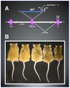
Figure 2
Epigenetic gene regulation at the Avy locus. (A) The Avy metastable epiallele contains an intracisternal A particle insertion within pseudoexon 1A. Normal transcription occurs from a hair cycle specific promoter in exon 2 and leads to brown mice. The IAP insertion upstream of the wild type promoter leads to constitutive expression of Agouti from the IAP cryptic promoter and yellow mice. Stochastic Methylation of CpG sites upstream of the cryptic promoter correlates inversely with Avy expression. (B) Fifteen-week old, genetically identical, Avy mice with varying coat colors. Yellow mice (left) are hypomethylated upstream of the Avy promoter while pseudoagouti mice (right) are hypermethylated at these CpG sites, recapitulating normal Agouti expression. Increasing levels of ectopic expression of Agouti in 15-week old Avy mice (from right to left) leads to obesity, tumorigenesis, and diabetes.
Figure 3
Figure 3
Effect of bisphenol A (BPA) and maternal dietary supplementation on the phenotype and epigenotype of Avy/a offspring. (A) Female mice were exposed to a modified control diet with corn oil substituted for soy bean oil, a modified diet containing 50 mg/kg BPA, or modified diets containing 50 mg/kg BPA and supplemented with 250 mg/kg genistein or methyl donors. (B) Offspring exposed to BPA in utero and during lactation were hypomethylated at the Avy allele and of a higher proportion yellow than control mice. Offspring that were exposed to BPA and supplemented with methyl donors and genistein returned to the methylation levels and coat color proportions of control.

References

    1. Anway M, Cupp A, Uzumcu M, Skinner M. Epigenetic transgenerational actions of endocrine disruptor and male fertility. Science. 2005;308(5727):1466–1469. - PMC - PubMed
    1. Anway MD, Skinner MK. Epigenetic programming of the germ line: effects of endocrine disruptors on the development of transgenerational disease. Reprod Biomed Online. 2008;16(1):23–25. - PMC - PubMed
    1. Baqir S, Smith L. Growth restricted in vitro culture conditions alter the imprinted gene expression patterns of mouse embryonic stem cells. Cloning Stem Cells. 2003;5(3):199–121. - PubMed
    1. Barker D, Eriksson J, Forsen T, Osmond C. Fetal origins of adult disease: Strength of effects and biological basis. Int J Epidemiol. 2002;31:1235–1239. - PubMed
    1. Barker D, Osmond C, Kajantie E, Eriksson J. Growth and chronic disease: findings in the Helsinki birth cohort. Annals of Human Biology. 2009;36(5):445–458. - PubMed

Publication types