. 2010 Sep;52(3):987-98.
doi: 10.1002/hep.23783.

Biliary apotopes and anti-mitochondrial antibodies activate innate immune responses in primary biliary cirrhosis

Affiliations

Biliary apotopes and anti-mitochondrial antibodies activate innate immune responses in primary biliary cirrhosis

Ana Lleo et al. Hepatology. 2010 Sep.

Abstract

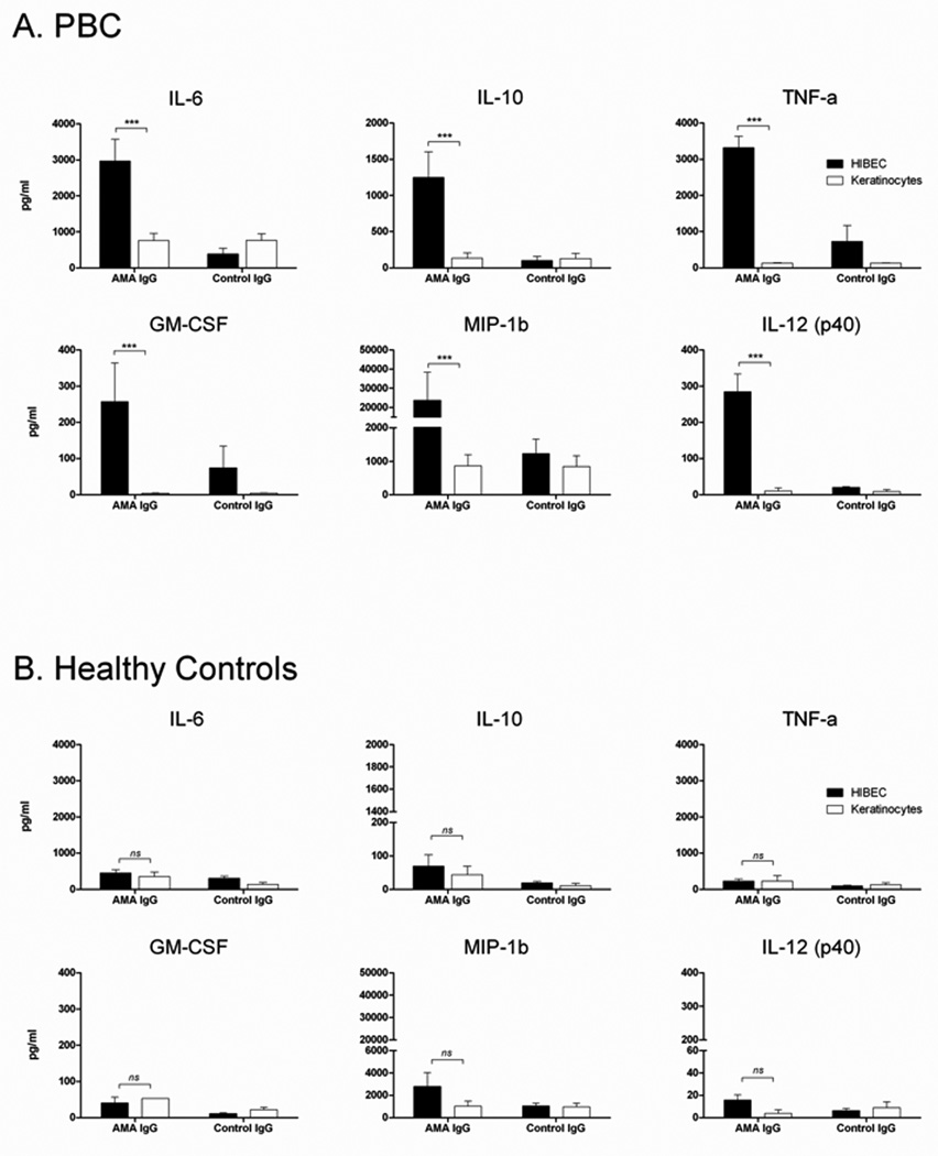
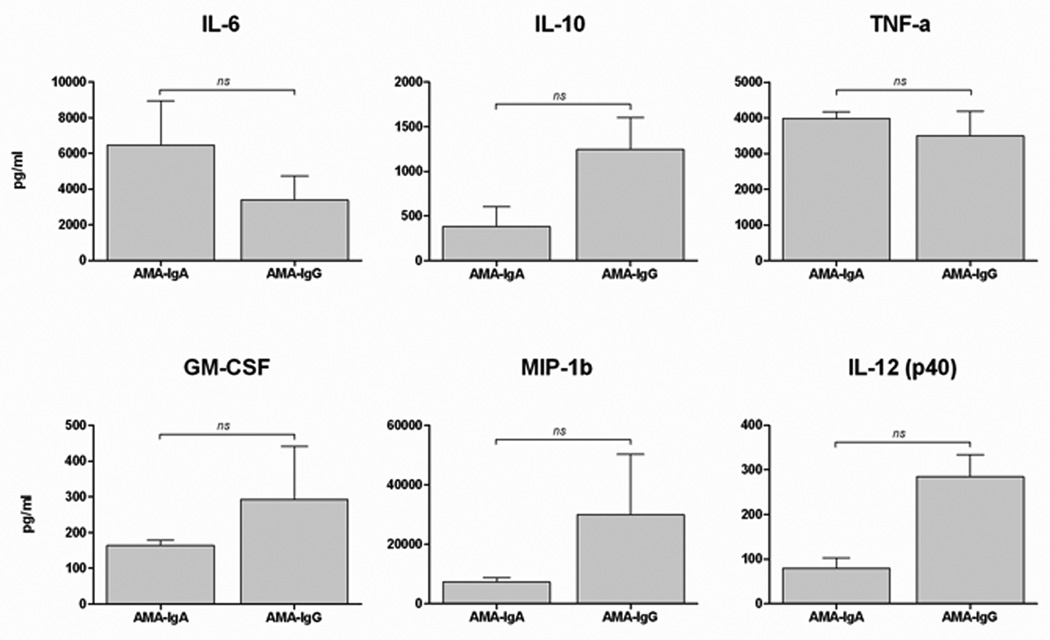
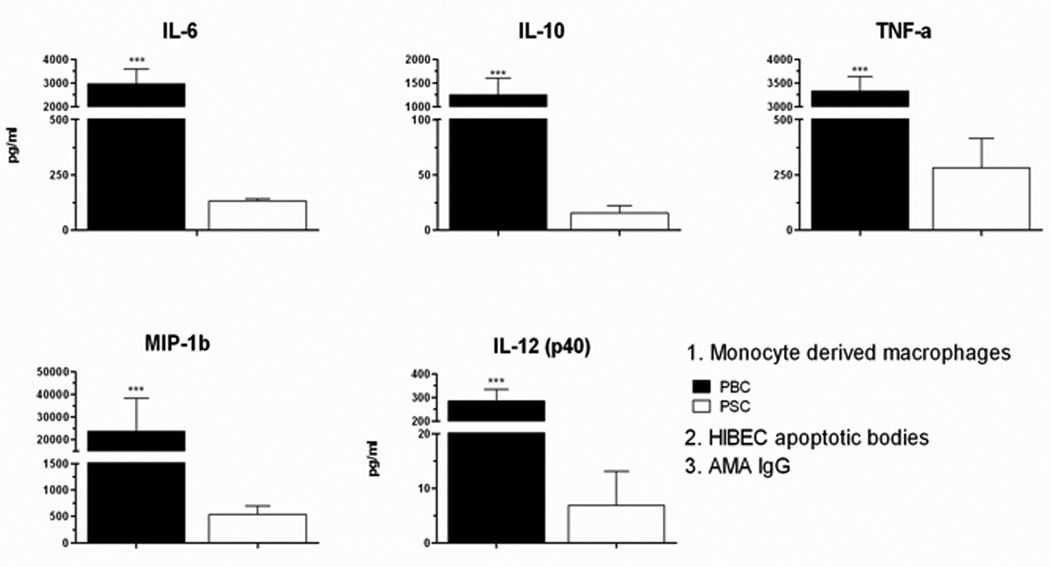
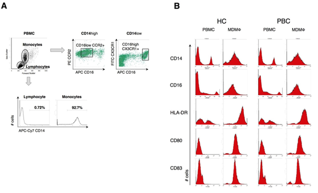
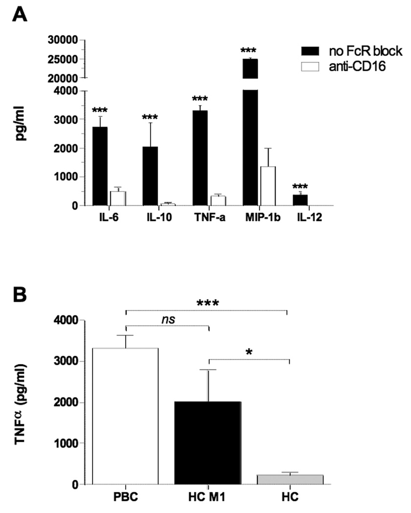
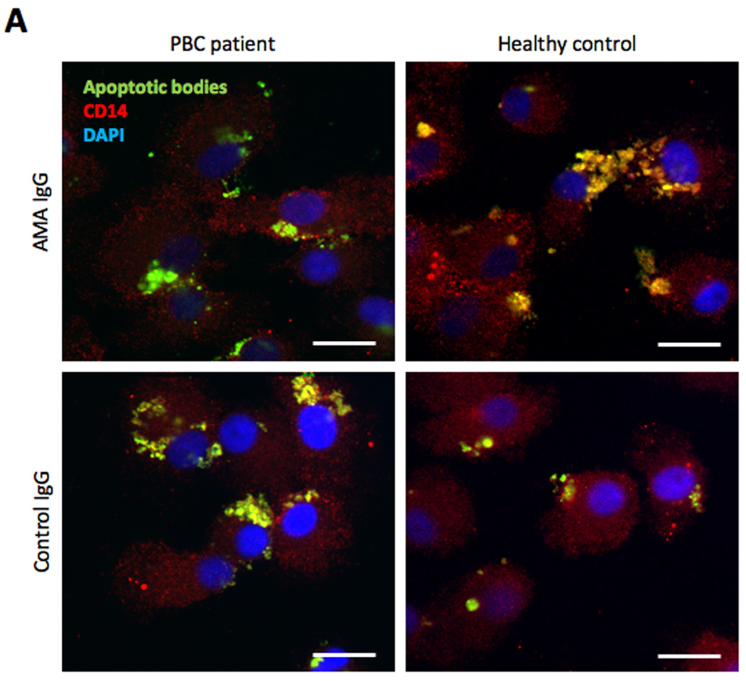
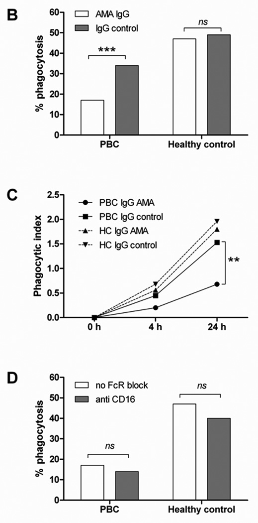
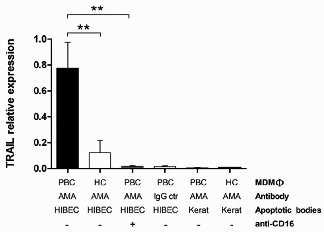
Our understanding of primary biliary cirrhosis (PBC) has been significantly enhanced by the rigorous dissection of the multilineage T and B cell response against the immunodominant mitochondrial autoantigen, the E2 component of the pyruvate dehydrogenase complex (PDC-E2). PDC-E2 is a ubiquitous protein present in mitochondria of nucleated cells. However, the damage of PBC is confined to small biliary epithelial cells (BECs). We have previously demonstrated that BECs translocate immunologically intact PDC-E2 to apoptotic bodies and create an apotope. To define the significance of this observation, we have studied the ability of biliary or control epithelial apotopes to induce cytokine secretion from mature monocyte-derived macrophages (MDMphis) from either patients with PBC or controls in the presence or absence of anti-mitochondrial antibodies (AMAs). We demonstrate that there is intense inflammatory cytokine production in the presence of the unique triad of BEC apotopes, macrophages from patients with PBC, and AMAs. The cytokine secretion is inhibited by anti-CD16 and is not due to differences in apotope uptake. Moreover, MDMphis from PBC patients cultured with BEC apoptotic bodies in the presence of AMAs markedly increase tumor necrosis factor-related apoptosis-inducing ligand expression.

Conclusion: These results provide a mechanism for the biliary specificity of PBC, the recurrence of disease after liver transplantation, and the success of ursodiol in treatment. They further emphasize the critical role of the innate immune system in the perpetuation of this autoimmune disease.


Conflict of interest statement

Conflict of interest: the authors declare that no conflict of interest exists

Figures

Figure 1
Figure 1. Macrophages from PBC cultured with apoptotic bodies from HIBEC secrete pro-inflammatory cytokines in the presence of AMA
MDMΦ from patients with PBC (A) or unaffected controls (B) were cultured with apoptotic bodies and antibody (AMA IgG or control IgG). The triad of MDMΦ from patients with PBC (A), but not from controls (B), apoptotic bodies from HIBEC cells, and AMA leads to significantly increased secretion of TNF-α, IL-6, IL-10, MIP-1b, and IL12p40 compared to IgG control. Each experiment was performed in duplicate. Level of significance is denoted as ***p<0.001, based on a two tailed Mann-Whitney test with 95% CI.
Figure 2
Figure 2. Both IgA and IgG isotypes stimulate MDMΦ from patients with PBC
MDMΦ from 10 patients with PBC and 10 controls were cultured with HIBEC apoptotic bodies in the presence of two different isotypes of AMA (IgG and IgA). Supernatants were collected after 24 h of incubation and cytokine concentration determined. While cytokine levels were markedly increased (see Figure 1A), there was no significant difference (ns) in levels of TNF-α, IL-6, Il-10, MIP-1b, IL12p40 and GM-CSF between AMA-IgG and IgA.
Figure 3
Figure 3. MDMΦ from PBC secrete significantly higher levels of cytokines compared to PSC patients
Cytokine secretion in supernatants collected at 24 hours from MDMΦ isolated from patients with PBC (n=25) or PSC (n=6) cultured with HIBEC apoptotic bodies in the presence of AMA. Levels of TNF-α, IL-6, Il-10, MIP-1b, and IL12p40 were significantly higher in cultures with MDMΦ from PBC compared to PSC. Statistical difference was analyzed by a two tailed Mann-Whitney test (95% CI), *** p < 0.001. All samples were run in duplicate.
Figure 4
Figure 4. Phenotype of circulating monocytes and MDMΦ as determined by FACS analysis of monocyte subsets and phenotype of monocytes and MDMΦ in patients with PBC and controls
Cells were stained with fluorochrome-conjugated antibodies to CD14, CD16, CCR2, CX3CR1, HLA-DR, CD80 and CD83. (A) Monocytes and lymphocytes from PBMCs were gated on the basis of forward and side scatter profiles and two monocyte subpopulations were identified: classical CD14highCD16- CCR2+ (87%) and a minor subset of CD14lowCD16+CX3CR1+ (13%). (B) The enriched population of CD14+ monocytes were stimulated to develop MDMΦ and cultured with apoptotic bodies. No differences in the phenotypes of PBMC and MDMΦ were observed between PBC and HC.
Figure 5
Figure 5. Stimulation of PBC macrophages in the presence of AMA and HIBEC apoptotic bodies is mediated by the Fc Receptor and is a consequence of M1 polarization
(A) Comparison of cytokine secretion from MDMΦ from patients with PBC (n=8) cultured with HIBEC apoptotic bodies in the presence of AMA with (white bars) or without (black bars) pre-treatment with CD16 monoclonal antibody (clone 3G8). Cytokine levels were markedly decreased when FcγR was blocked (***p< 0.001). (B) Comparison of TNFα secretion from MDMΦ from patients with PBC (n = 8) and healthy controls (n = 8) after M1 polarization (HC M1), and untreated (HC) cultured with HIBEC apoptotic bodies in the presence of AMA. (***p< 0.001, *p<0.05).
Figure 6
Figure 6. Uptake of apoptotic bodies
(A) Confocal imaging of MDMΦ from a representative patient with PBC and an unaffected control co-incubated for 24 h with CFSE-labeled apoptotic bodies from HIBEC in the presence of human AMA-IgG or control- IgG. After culture the cells were fixed and stained with PE-conjugated anti-human CD14 (red), DAPI was used to stain the nucleus (blue), CFSE stained apoptotic bodies are shown in green. In all four conditions, apoptotic bodies (green) are located inside the cells, which indicate that MDMΦ actively engulfed apoptotic bodies after 24 h (arrows). Scale bar 20 µm. (B) Comparison of phagocytic efficacy, expressed as percentage of phagocytosis, of MDMΦ from PBC and controls; MDMΦ from PBC had a reduced uptake of HIBEC apoptotic bodies in the presence of AMA (17% vs 47%, ***p<0.001), this difference was not noted when IgG control was used (34% vs 49%, p = ns). Percentage of phagocytosis was calculated by counting the number of macrophages that had ingested at least one HIBEC apoptotic body. (Fisher's Exact Test). (C) The phagocytic index (PI) was expressed as percentage of phagocytosis multiplied by the mean number of phagocytosed bodies per macrophage (ABMΦ): PI = (% phagocytosis x mean ABMΦ/100) and was evaluated at 0, 4 and 24 h of incubation. ** p< 0.01 (D) The ability of MDMΦ from PBC and controls to uptake HIBEC apoptotic bodies, expressed as percentage of phagocytosis, was studied in presence of AMA, with or without FcγR block. No significant difference (ns) in the percentage of phagocytosis was observed when FcγR was blocked in both patients and controls. Statistical difference was determined by Fisher's Exact Test.
Figure 7
Figure 7. TRAIL expression is increased in MDMΦ from PBC patients cultured with apoptotic bodies from HIBEC in the presence of AMA
The relative expression levels of TRAIL in MDMΦ are compared with regards to MDMΦ, AMA, cell type of apoptotic bodies, and use of anti-CD16. Note that TRAIL expression is increased at least 4 fold (**p < 0.01) in PBC MDMΦ, AMA and apoptotic bodies from HIBEC and significantly reduced (**p < 0.01) when the cells are pretreated with anti-CD16.

References

    1. Kaplan MM, Gershwin ME. Primary biliary cirrhosis. N Engl J Med. 2005;353:1261–1273. - PubMed
    1. Kawano A, Shimoda S, Kamihira T, Ishikawa F, Niiro H, Soejima Y, Taketomi A, et al. Peripheral tolerance and the qualitative characteristics of autoreactive T cell clones in primary biliary cirrhosis. J Immunol. 2007;179:3315–3324. - PubMed
    1. Shimoda S, Nakamura M, Ishibashi H, Hayashida K, Niho Y. HLA DRB4 0101-restricted immunodominant T cell autoepitope of pyruvate dehydrogenase complex in primary biliary cirrhosis: evidence of molecular mimicry in human autoimmune diseases. J Exp Med. 1995;181:1835–1845. - PMC - PubMed
    1. Shimoda S, Miyakawa H, Nakamura M, Ishibashi H, Kikuchi K, Kita H, Niiro H, et al. CD4 T-cell autoreactivity to the mitochondrial autoantigen PDC-E2 in AMA-negative primary biliary cirrhosis. J Autoimmun. 2008;31:110–115. - PubMed
    1. Yang GX, Lian ZX, Chuang YH, Moritoki Y, Lan RY, Wakabayashi K, Ansari AA, et al. Adoptive transfer of CD8(+) T cells from transforming growth factor beta receptor type II (dominant negative form) induces autoimmune cholangitis in mice. Hepatology. 2008;47:1974–1982. - PMC - PubMed

Publication types

MeSH terms