

Sulfur mustard vapor effects on differentiated human lung cells
- PMID: 20569120
- PMCID: PMC3214633
- DOI: 10.3109/08958378.2010.493901
Sulfur mustard vapor effects on differentiated human lung cells
Abstract
Context: Sulfur mustard (SM) causes skin blistering and long-term pulmonary dysfunction. Its adverse effects have been studied in battlefield-exposed humans, but lack of knowledge regarding confounding factors makes interpretation challenging. Animal studies are critical to understanding mechanisms, but differences between animals and humans must be addressed. Studies of cultured human cells can bridge animal studies and humans.
Objective: Evaluate effects of SM vapor on airway cells.
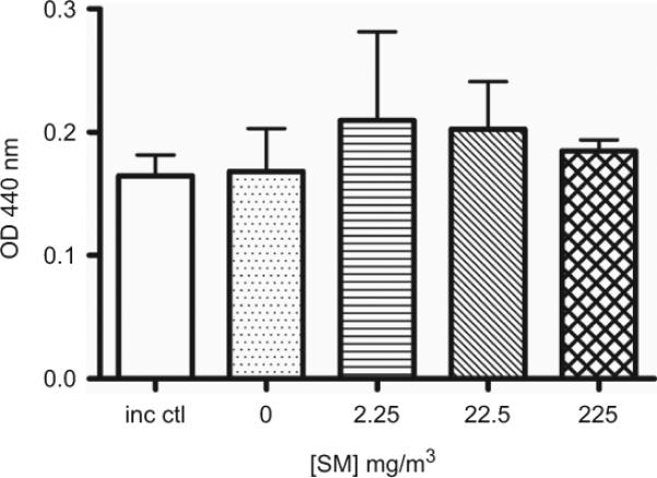
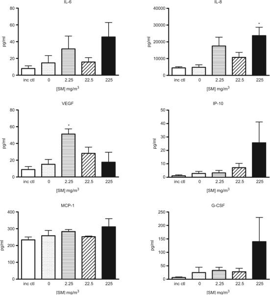
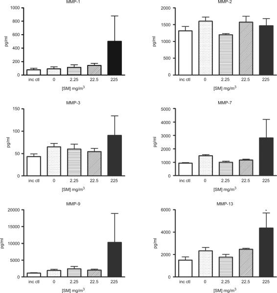
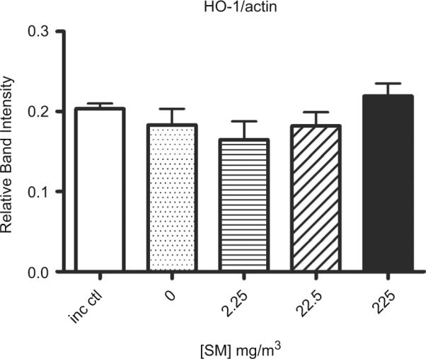
Materials and methods: We examined responses of differentiated human tracheal/bronchial epithelial cells, cultured at an air-liquid interface, to SM vapors. SM effects on metabolic activity (Water Soluble Tetrazolium (WST) assay), cytokine and metalloproteinase secretion, and cellular heme oxygenase 1 (HO-1), an oxidative stress indicator, were measured after 24 h.
Results: At noncytotoxic levels of exposure, interleukin 8 and matrix metalloproteinase-13 were significantly increased in these cultures, but HO-1 was not significantly affected.
Discussion and conclusion: Exposure of differentiated airway epithelial cells to sub-cytotoxic levels of SM vapor induced inflammatory and degradative responses that could contribute to the adverse health effects of inhaled SM.
Figures
Similar articles
-
Suppression of inducible nitric oxide synthase expression and nitric oxide production by macrolide antibiotics in sulfur mustard-exposed airway epithelial cells.Basic Clin Pharmacol Toxicol. 2008 Sep;103(3):255-61. doi: 10.1111/j.1742-7843.2008.00255.x. Epub 2008 Jul 18. Basic Clin Pharmacol Toxicol. 2008. PMID: 18684233
-
Pulmonary injury and oxidative stress in rats induced by inhaled sulfur mustard is ameliorated by anti-tumor necrosis factor-α antibody.Toxicol Appl Pharmacol. 2021 Oct 1;428:115677. doi: 10.1016/j.taap.2021.115677. Epub 2021 Aug 11. Toxicol Appl Pharmacol. 2021. PMID: 34390737 Free PMC article.
-
Inhibition of sulfur mustard-induced cytotoxicity and inflammation by the macrolide antibiotic roxithromycin in human respiratory epithelial cells.BMC Cell Biol. 2007 May 24;8:17. doi: 10.1186/1471-2121-8-17. BMC Cell Biol. 2007. PMID: 17524151 Free PMC article.
-
Respiratory effects of sulfur mustard exposure, similarities and differences with asthma and COPD.Inhal Toxicol. 2015;27(14):731-44. doi: 10.3109/08958378.2015.1114056. Epub 2015 Dec 4. Inhal Toxicol. 2015. PMID: 26635274 Review.
-
Acute and chronic effects of sulfur mustard on the skin: a comprehensive review.Cutan Ocul Toxicol. 2010 Dec;29(4):269-77. doi: 10.3109/15569527.2010.511367. Epub 2010 Sep 24. Cutan Ocul Toxicol. 2010. PMID: 20868209 Review.
Cited by
-
Sulfur mustard-induced pulmonary injury: therapeutic approaches to mitigating toxicity.Pulm Pharmacol Ther. 2011 Feb;24(1):92-9. doi: 10.1016/j.pupt.2010.09.004. Epub 2010 Sep 17. Pulm Pharmacol Ther. 2011. PMID: 20851203 Free PMC article. Review.
-
Inhalation of sulfur mustard causes long-term T cell-dependent inflammation: possible role of Th17 cells in chronic lung pathology.Int Immunopharmacol. 2012 May;13(1):101-8. doi: 10.1016/j.intimp.2012.03.010. Epub 2012 Mar 28. Int Immunopharmacol. 2012. PMID: 22465472 Free PMC article.
-
From the Cover: Catalytic Antioxidant Rescue of Inhaled Sulfur Mustard Toxicity.Toxicol Sci. 2016 Dec;154(2):341-353. doi: 10.1093/toxsci/kfw170. Epub 2016 Sep 7. Toxicol Sci. 2016. PMID: 27605419 Free PMC article.
-
Invited review: human air-liquid-interface organotypic airway tissue models derived from primary tracheobronchial epithelial cells-overview and perspectives.In Vitro Cell Dev Biol Anim. 2021 Feb;57(2):104-132. doi: 10.1007/s11626-020-00517-7. Epub 2020 Nov 11. In Vitro Cell Dev Biol Anim. 2021. PMID: 33175307 Free PMC article. Review.
-
Assessment of biological responses of EpiAirway 3-D cell constructs versus A549 cells for determining toxicity of ambient air pollution.Inhal Toxicol. 2016;28(6):251-9. doi: 10.3109/08958378.2016.1157227. Inhal Toxicol. 2016. PMID: 27100558 Free PMC article.
References
-
- Anderson DR, Byers SL, Clark CR, Schleicher E. Biochemical alterations in rat lung lavage fluid following acute sulfur mustard inhalation. Inhalation Toxicology. 1996;9:43–51.
-
- Anderson DR, Yourick JJ, Moeller RB, Petrali JP, Young GD, Byers SL. Pathologic changes in rat lungs following acute sulfur mustard inhalation. Inhalation Toxicology. 1997;8:285–297.
-
- Arroyo CM, Broomfield CA, Hackley BE., Jr. The role of interleukin-6 (IL-6) in human sulfur mustard (HD) toxicology. Int J Toxicol. 2001;20:281–296. - PubMed
-
- Arroyo CM, Schafer RJ, Kurt EM, Broomfield CA, Carmichael AJ. Response of normal human keratinocytes to sulfur mustard (HD): Cytokine release using a non-enzymatic detachment procedure. Hum Exp Toxicol. 1999;18:1–11. - PubMed
-
- Arroyo CM, Schafer RJ, Kurt EM, Broomfield CA, Carmichael AJ. Response of normal human keratinocytes to sulfur mustard: Cytokine release. J Appl Toxicol. 2000;20(Suppl 1):S63–S72. - PubMed
Publication types
MeSH terms
Substances
Grants and funding
LinkOut - more resources
Full Text Sources