. 2010 Oct;9(5):911-5.
doi: 10.1111/j.1474-9726.2010.00598.x.

Tendon-derived stem/progenitor cell aging: defective self-renewal and altered fate

Affiliations

Tendon-derived stem/progenitor cell aging: defective self-renewal and altered fate

Zuping Zhou et al. Aging Cell. 2010 Oct.

Abstract

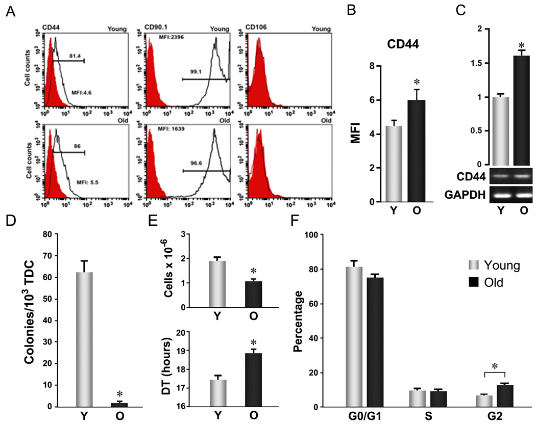
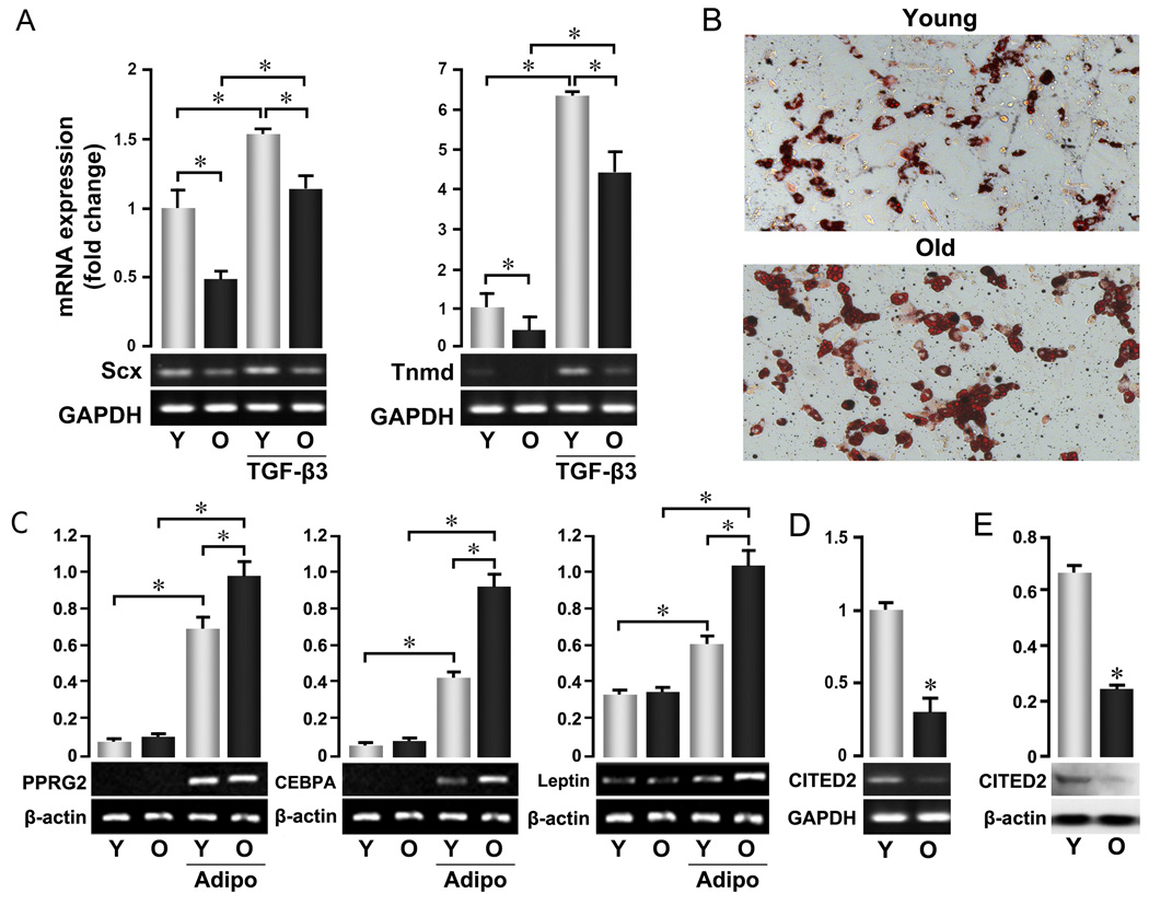
Aging is a major risk factor for tendon injury and impaired tendon healing, but the basis for these relationships remains poorly understood. Here we show that rat tendon- derived stem ⁄ progenitor cells (TSPCs) differ in both self-renewal and differentiation capability with age. The frequency of TSPCs in tendon tissues of aged animals is markedly reduced based on colony formation assays. Proliferation rate is decreased, cell cycle progression is delayed and cell fate patterns are also altered in aged TSPCs. In particular, expression of tendon lineage marker genes is reduced while adipocytic differentiation increased. Cited2, a multi-stimuli responsive transactivator involved in cell growth and senescence, is also downregulated in aged TSPCs while CD44, a matrix assembling and organizing protein implicated in tendon healing, is upregulated, suggesting that these genes participate in the control of TSPC function.


Figures

Figure 1
Figure 1. Aging-related cellular changes in TSPC self-renewal
Young and aged TSPCs were prepared, as described by Bi et al (Bi et al. 2007), from 3–4 and 24–26 month Sprague-Dawley male rats, respectively. Patellar tendons were digested with collagenase A/dispase (2 h, 37 °C) and cultured in DMEM plus 10% FBS for 7–9 days. Adherent cells (passage 0, P0) at the end of culture were used as TSPCs for all assays unless otherwise specified. (A–C) Age-related surface antigen expression changes in TSPCs. A: Representative histogram plots. TSPCs stained with fluorescein-conjugated anti-rat antibodies (open) or isotype control antibodies (filled) were analyzed by FACS. B: Mean fluorescence intensity of CD44 based on FACS analysis of surface expression as shown in A (n=3, independent experiments, *P<0.05). C: CD44 mRNA levels determined by RT-PCR. Upper Panels represent the quantification of band intensity from the gel shown in lower panels. (D) Colony formation. Total tendon-derived cells (TDC) were plated at 1×10³ cells/well (6-well plate) and grown for 9 days. Colony forming units (CFUs) were scored after methylene blue staining. Error bars represent SD (n=3, *P<0.05 versus aged TSPC). (E) Proliferation. TSPCs were seeded at 1×10³ cells/well (6 well plate) and cultured for 8 days. Cell numbers were counted and cell population doubling times were estimated from start (0) and end (1) points. Error bars represent SD (n=3, *P<0.05 versus aged TSPC). (F) Cell cycle distribution. TSPCs were fixed with 70% ethanol, stained with propidium iodide (PI), and analyzed by FACS. Error bars represent SD (n=3 independent experiments; * P<0.05)
Figure 2
Figure 2. Altered cell fate of aged TSPCs
P0 TSPCs were prepared as described in Figure 1 and used for all assays. (A) Expression of tendon lineage-specific genes. TSPCs were cultured with or without TGF-β3 for 3 days, and total RNA was extracted after culture. (B, C) Adipocyte-skewed differentiation of aged TSPCs. Cells were cultured in specific induction medium for 16 days. B: Oil Red O staining per (Gimble et al. 1995); C: mRNA expression of adipogenic marker genes in untreated and adipogenic-induced TSPCs. (D, E) Expression of Cited2 at mRNA (D) and protein (E) levels in young and old TSPCs. mRNA expression of indicated genes in A, C, D was assessed by RT-PCR. Upper Panels represent the quantification of band intensity from the gel shown in lower panels. Data are representative of 3 experiments and confirmed by real-time PCR.

Similar articles

Cited by

References

    1. Ansorge HL, Beredjiklian PK, Soslowsky LJ. CD44 deficiency improves healing tendon mechanics and increases matrix and cytokine expression in a mouse patellar tendon injury model. J Orthop Res. 2009;27:1386–1391. - PMC - PubMed
    1. Bi Y, Ehirchiou D, Kilts TM, Inkson CA, Embree MC, Sonoyama W, Li L, Leet AI, Seo BM, Zhang L, Shi S, Young MF. Identification of tendon stem/progenitor cells and the role of the extracellular matrix in their niche. Nat Med. 2007;13:1219–1227. - PubMed
    1. Birch HL, Bailey JV, Bailey AJ, Goodship AE. Age-related changes to the molecular and cellular components of equine flexor tendons. Equine Vet J. 1999;31:391–396. - PubMed
    1. Chen Y, Haviernik P, Bunting KD, Yang YC. Cited2 is required for normal hematopoiesis in the murine fetal liver. Blood. 2007;110:2889–2898. - PMC - PubMed
    1. Couppe C, Hansen P, Kongsgaard M, Kovanen V, Suetta C, Aagaard P, Kjaer M, Magnusson SP. Mechanical properties and collagen cross-linking of the patellar tendon in old and young men. J Appl Physiol. 2009;107:880–886. - PubMed

Publication types