The role of iron uptake in pathogenicity and symbiosis in Photorhabdus luminescens TT01
- PMID: 20569430
- PMCID: PMC2905363
- DOI: 10.1186/1471-2180-10-177
The role of iron uptake in pathogenicity and symbiosis in Photorhabdus luminescens TT01
Abstract
Background: Photorhabdus are Gram negative bacteria that are pathogenic to insect larvae whilst also having a mutualistic interaction with nematodes from the family Heterorhabditis. Iron is an essential nutrient and bacteria have different mechanisms for obtaining both the ferrous (Fe2+) and ferric (Fe3+) forms of this metal from their environments. In this study we were interested in analyzing the role of Fe3+ and Fe2+ iron uptake systems in the ability of Photorhabdus to interact with its invertebrate hosts.
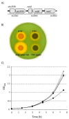
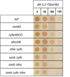
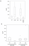
Results: We constructed targeted deletion mutants of exbD, feoABC and yfeABCD in P. luminescens TT01. The exbD mutant was predicted to be crippled in its ability to obtain Fe3+ and we show that this mutant does not grow well in iron-limited media. We also show that this mutant was avirulent to the insect but was unaffected in its symbiotic interaction with Heterorhabditis. Furthermore we show that a mutation in feoABC (encoding a predicted Fe2+ permease) was unaffected in both virulence and symbiosis whilst the divalent cation transporter encoded by yfeABCD is required for virulence in the Tobacco Hornworm, Manduca sexta (Lepidoptera) but not in the Greater Wax Moth, Galleria mellonella (Lepidoptera). Moreover the Yfe transporter also appears to have a role during colonization of the IJ stage of the nematode.
Conclusion: In this study we show that iron uptake (via the TonB complex and the Yfe transporter) is important for the virulence of P. luminescens to insect larvae. Moreover this study also reveals that the Yfe transporter appears to be involved in Mn2+-uptake during growth in the gut lumen of the IJ nematode. Therefore, the Yfe transporter in P. luminescens TT01 is important during colonization of both the insect and nematode and, moreover, the metal ion transported by this pathway is host-dependent.
Figures


Similar articles
-
The pbgPE operon in Photorhabdus luminescens is required for pathogenicity and symbiosis.J Bacteriol. 2005 Jan;187(1):77-84. doi: 10.1128/JB.187.1.77-84.2005. J Bacteriol. 2005. PMID: 15601690 Free PMC article.
-
The exbD gene of Photorhabdus temperata is required for full virulence in insects and symbiosis with the nematode Heterorhabditis.Mol Microbiol. 2005 May;56(3):763-73. doi: 10.1111/j.1365-2958.2005.04574.x. Mol Microbiol. 2005. PMID: 15819630
-
Photorhabdus luminescens genes induced upon insect infection.BMC Genomics. 2008 May 19;9:229. doi: 10.1186/1471-2164-9-229. BMC Genomics. 2008. PMID: 18489737 Free PMC article.
-
The genetic basis of the symbiosis between Photorhabdus and its invertebrate hosts.Adv Appl Microbiol. 2014;88:1-29. doi: 10.1016/B978-0-12-800260-5.00001-2. Adv Appl Microbiol. 2014. PMID: 24767424 Review.
-
Comparative analysis of the Photorhabdus luminescens and the Yersinia enterocolitica genomes: uncovering candidate genes involved in insect pathogenicity.BMC Genomics. 2008 Jan 25;9:40. doi: 10.1186/1471-2164-9-40. BMC Genomics. 2008. PMID: 18221513 Free PMC article. Review.
Cited by
-
Preliminary Screening on Antibacterial Crude Secondary Metabolites Extracted from Bacterial Symbionts and Identification of Functional Bioactive Compounds by FTIR, HPLC and Gas Chromatography-Mass Spectrometry.Molecules. 2024 Jun 19;29(12):2914. doi: 10.3390/molecules29122914. Molecules. 2024. PMID: 38930979 Free PMC article.
-
Pathogenomics of Virulence Traits of Plesiomonas shigelloides That Were Deemed Inconclusive by Traditional Experimental Approaches.Front Microbiol. 2018 Dec 21;9:3077. doi: 10.3389/fmicb.2018.03077. eCollection 2018. Front Microbiol. 2018. PMID: 30627119 Free PMC article.
-
Aerobactin synthesis genes iucA and iucC contribute to the pathogenicity of avian pathogenic Escherichia coli O2 strain E058.PLoS One. 2013;8(2):e57794. doi: 10.1371/journal.pone.0057794. Epub 2013 Feb 27. PLoS One. 2013. PMID: 23460907 Free PMC article.
-
Commensal Pseudomonas protect Arabidopsis thaliana from a coexisting pathogen via multiple lineage-dependent mechanisms.ISME J. 2022 May;16(5):1235-1244. doi: 10.1038/s41396-021-01168-6. Epub 2021 Dec 11. ISME J. 2022. PMID: 34897280 Free PMC article.
-
Toward a better understanding of the mechanisms of symbiosis: a comprehensive proteome map of a nascent insect symbiont.PeerJ. 2017 May 9;5:e3291. doi: 10.7717/peerj.3291. eCollection 2017. PeerJ. 2017. PMID: 28503376 Free PMC article.
References
-
- Clarke DJ, Dowds BCA. Virulence mechanisms of Photorhabdus sp strain K122 toward wax moth larvae. J Invert Pathol. 1995;66:149–155. doi: 10.1006/jipa.1995.1078. - DOI
Publication types
MeSH terms
Substances
Grants and funding
LinkOut - more resources
Full Text Sources
Medical
Molecular Biology Databases

