. 2010 Jun 22:9:157.
doi: 10.1186/1476-4598-9-157.

Nerve growth factor promotes breast cancer angiogenesis by activating multiple pathways

Affiliations

Nerve growth factor promotes breast cancer angiogenesis by activating multiple pathways

Rodrigue Romon et al. Mol Cancer. .

Abstract

Background: Although several anti-angiogenic therapies have been approved in the treatment of cancer, the survival benefits of such therapies are relatively modest. Discovering new molecules and/or better understating signaling pathways of angiogenesis is therefore essential for therapeutic improvements. The objective of the present study was to determine the involvement of nerve growth factor (NGF) in breast cancer angiogenesis and the underlying molecular mechanisms.

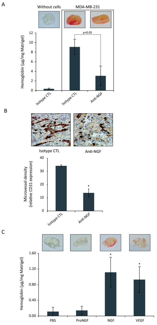
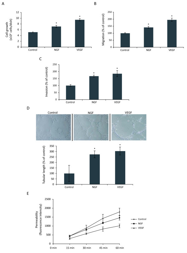
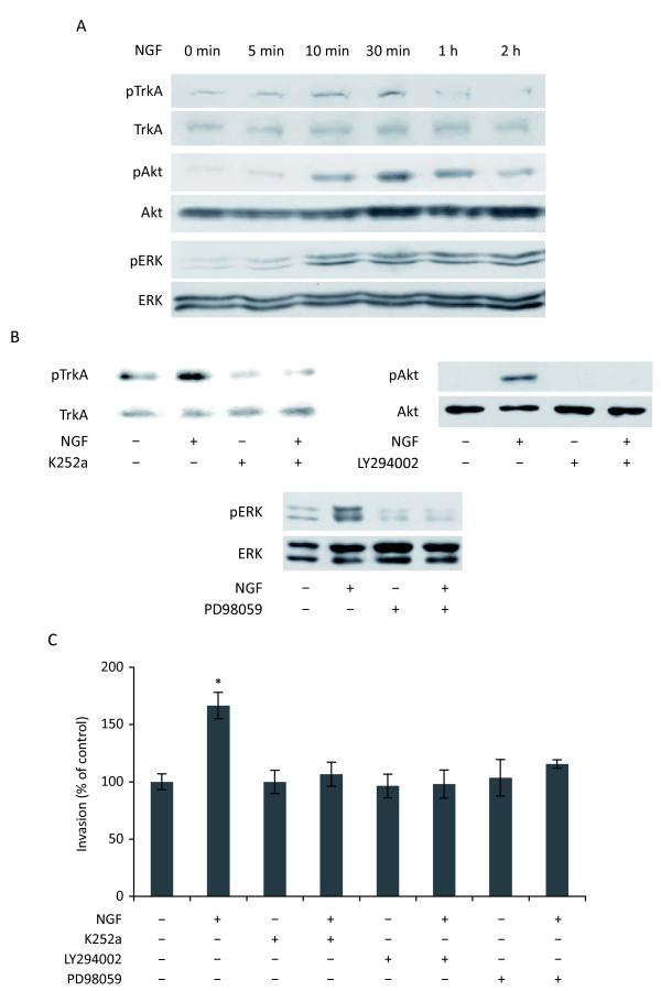
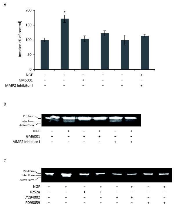
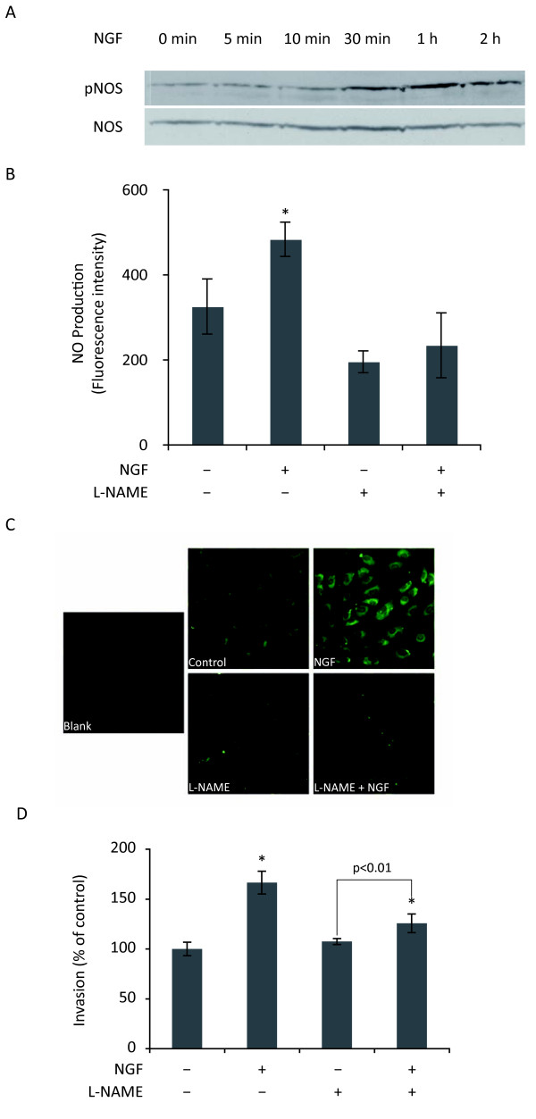
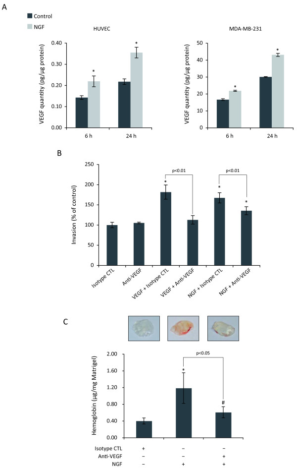
Results: We showed that both recombinant NGF and NGF produced by breast cancer cells stimulated angiogenesis in Matrigel plugs in immunodeficient mice. NGF strongly increased invasion, cord formation and the monolayer permeability of endothelial cells. Moreover, NGF-stimulated invasion was under the control of its tyrosine kinase receptor (TrkA) and downstream signaling pathways such as PI3K and ERK, leading to the activation of matrix metalloprotease 2 and nitric oxide synthase. Interestingly, NGF increased the secretion of VEGF in both endothelial and breast cancer cells. Inhibition of VEGF, with a neutralizing antibody, reduced about half of NGF-induced endothelial cell invasion and angiogenesis in vivo.

Conclusions: Our findings provided direct evidence that NGF could be an important stimulator for breast cancer angiogenesis. Thus, NGF, as well as the activated signaling pathways, should be regarded as potential new targets for anti-angiogenic therapy against breast cancer.


Figures

Figure 1
Figure 1
Angiogenesis assay using Matrigel plugs in SCID mice. Matrigel containing different reagents was subcutaneously injected into SCID mice as described in materials and methods. (A, B) Matrigel was mixed with MDA-MB-231 breast cancer cells and isotype control or anti-NGF neutralizing antibodies (75 μg/ml); (C) Matrigel was mixed with proNGF (7.5 μg/ml), NGF (3.75 μg/ml), or VEGF (0.375 μg/ml). Angiogenesis was analyzed by quantification of hemoglobin (A, C) and microvessel density (B) as described in materials and methods. Five mice were used for each group and results are the mean of three independent experiments. Student's t-test, *p < 0.01 versus control.
Figure 2
Figure 2
Pleiotropic effects of NGF on HUVEC. (A) Growth assay. HUVEC cultured on standard culture plastic were treated with NGF (100 ng/ml), or VEGF (10 ng/ml) for 48 h. (B and C) Migration and invasion assay using Transwells. (D) Cord formation assay on matrigel-coated 24-well dishes. (E) Endothelial cell monolayer permeability assay. The kinetics of 70 kDa Dextran-FITC having passed through the monolayer of endothelial cells was determined by measurement of fluorescence in the lower chambers at different time points. A to D Student's t-test, *p < 0.01 versus control. E, ANOVA test, p < 0.05.
Figure 3
Figure 3
Involvement of TrkA, PI3K/Akt and MAPK pathways in NGF-stimulated invasion of HUVEC. (A) Western blot analysis of phospho TrkA (pTrkA), pAkt, and pERK upon NGF (100 ng/ml) stimulation. (B) Inhibition of pTrkA, pAkt and pERK by specific pharmacological inhibitors. HUVEC were pretreated with K252a, LY294002 or PD98059 for 30 min before treatment with NGF for another 20 min. For both A and B, results are representative of at least two independent experiments. (C) Invasion assay using Transwells. HUVEC were pretreated with the indicated pharmacological inhibitors for 30 min and then treated with NGF for 24 h. Student's t-test, *p < 0.01 versus control.
Figure 4
Figure 4
Involvement of MMPs in NGF-stimulated invasion. (A) Invasion assay using Transwells. HUVEC were pretreated with the broad spectrum inhibitor of MMPs GM6001 (10 μM) or the specific inhibitor of MMP2 (MMP2 Inhibitor I, 5 μM) for 30 min and then treated with NGF (100 ng/ml) for 24 h. Student's t-test, *p < 0.01 versus control. (B and C) Zymography analysis of MMP2. HUVEC were treated with NGF in the presence of different pharmacological inhibitors as described above before zymography analysis. Results are representative of two independent experiments.
Figure 5
Figure 5
Involvement of NO synthase in NGF-stimulated invasion. (A) Western blot analysis of phospho NO Synthase (pNOS) upon NGF (100 ng/ml) treatment. Results were representative of two independent experiments. (B) Quantification of NO levels in HUVEC. Cells were pretreated with NO synthase inhibitor (L-NAME, 0.1 mM) for 30 min, then loaded with DAF-2DA for 20 min before treatment with NGF in the presence or absence of L-NAME for 2 h. (C) Illustration of fluorescence intensity, which represent the levels of NO in HUVEC under different conditions. (D) Invasion assay using Transwells. HUVEC were pretreated with L-NAME and then treated with NGF for 24 h. For both B and D, Student's t-test, *p < 0.01 versus control.
Figure 6
Figure 6
Involvement of the VEGF in NGF-stimulated angiogenesis. (A) ELISA quantification of VEGF in conditioned media from HUVEC and MDA-MB-231 cells. Cells were treated with NGF (100 ng/ml) for 6 h or 24 h, conditioned media were concentrated before ELISA assay, as described in materials and methods. (B) Invasion assay using Transwells. HUVEC were treated with NGF (100 ng/ml) or VEGF (10 ng/ml) in the presence of isotype control or anti-VEGF neutralizing antibodies (1 μg/ml) for 24 h. (C) In vivo angiogenesis assay. Matrigel containing a mixture of NGF and isotype control or anti-VEGF neutralizing antibodies (37.5 μg/ml) was subcutaneously injected into SCID mice (five mice per group) as described in materials and methods. Hemoglobin in Matrigel plugs was quantified by Drabkin method 7 days after injection. Results are the mean of three independent experiments. Student's t-test, *p < 0.01; #p < 0.05 versus control.

References

    1. Folkman J, Watson K, Ingber D, Hanahan D. Induction of angiogenesis during the transition from hyperplasia to neoplasia. Nature. 1989;339:58–61. - PubMed
    1. Furstenberger G, von Moos R, Lucas R, Thurlimann B, Senn HJ, Hamacher J, Boneberg EM. Circulating endothelial cells and angiogenic serum factors during neoadjuvant chemotherapy of primary breast cancer. Br J Cancer. 2006;94:524–531. - PMC - PubMed
    1. Hicklin DJ, Ellis LM. Role of the vascular endothelial growth factor pathway in tumor growth and angiogenesis. J Clin Oncol. 2005;23:1011–1027. - PubMed
    1. Le Bourhis X, Romon R, Hondermarck H. Role of endothelial progenitor cells in breast cancer angiogenesis: from fundamental research to clinical ramifications. Breast Cancer Res Treat. 2010;120:17–24. - PubMed
    1. Kim KJ, Li B, Winer J, Armanini M, Gillett N, Phillips HS, Ferrara N. Inhibition of vascular endothelial growth factor-induced angiogenesis suppresses tumour growth in vivo. Nature. 1993;362:841–844. - PubMed

Publication types