Radiobiological evaluation of forward and inverse IMRT using different fractionations for head and neck tumours
- PMID: 20569482
- PMCID: PMC2907388
- DOI: 10.1186/1748-717X-5-57
Radiobiological evaluation of forward and inverse IMRT using different fractionations for head and neck tumours
Abstract
Purpose: To quantify the radiobiological advantages obtained by an Improved Forward Planning technique (IFP) and two IMRT techniques using different fractionation schemes for the irradiation of head and neck tumours. The conventional radiation therapy technique (CONVT) was used here as a benchmark.
Methods: Seven patients with head and neck tumours were selected for this retrospective planning study. The PTV1 included the primary tumour, PTV2 the high risk lymph nodes and PTV3 the low risk lymph nodes. Except for the conventional technique where a maximum dose of 64.8 Gy was prescribed to the PTV1, 70.2 Gy, 59.4 Gy and 50.4 Gy were prescribed respectively to PTV1, PTV2 and PTV3. Except for IMRT2, all techniques were delivered by three sequential phases. The IFP technique used five to seven directions with a total of 15 to 21 beams. The IMRT techniques used five to nine directions and around 80 segments. The first, IMRT1, was prescribed with the conventional fractionation scheme of 1.8 Gy per fraction delivered in 39 fractions by three treatment phases. The second, IMRT2, simultaneously irradiated the PTV2 and PTV3 with 59.4 Gy and 50.4 Gy in 28 fractions, respectively, while the PTV1 was boosted with six subsequent fractions of 1.8 Gy. Tissue response was calculated using the relative seriality model and the Poisson Linear-Quadratic-Time model to simulate repopulation in the primary tumour.
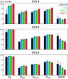
Results: The average probability of total tumour control increased from 38% with CONVT to 80% with IFP, to 85% with IMRT1 and 89% with IMRT2. The shorter treatment time and larger dose per fraction obtained with IMRT2 resulted in an 11% increase in the probability of control in the PTV1 with respect to IFP and 7% relatively to IMRT1 (p < 0.05). The average probability of total patient complications was reduced from 80% with CONVT to 61% with IFP and 31% with IMRT. The corresponding probability of complications in the ipsilateral parotid was 63%, 42% and 20%; in the contralateral parotid it was 50%, 20% and 9%; in the oral cavity it was 2%, 15% and 4% and in the mandible it was 1%, 5% and 3%, respectively.
Conclusions: A significant improvement in treatment outcome was obtained with IMRT compared to conventional radiation therapy. The practical and biological advantages of IMRT2, employing a shorter treatment time, may outweigh the small differences obtained in the organs at risk between the two IMRT techniques. This technique is therefore presently being used in the clinic for selected patients with head and neck tumours. A significant improvement in the quality of the dose distribution was obtained with IFP compared to CONVT. Thus, this beam arrangement is used in the clinical routine as an alternative to IMRT.
Figures


Similar articles
-
Significant improvement in normal tissue sparing and target coverage for head and neck cancer by means of helical tomotherapy.Radiother Oncol. 2006 Mar;78(3):276-82. doi: 10.1016/j.radonc.2006.02.009. Epub 2006 Mar 20. Radiother Oncol. 2006. PMID: 16546279 Clinical Trial.
-
Comparing 3DCRT and inversely optimized IMRT planning for head and neck cancer: equivalence between step-and-shoot and sliding window techniques.Radiother Oncol. 2005 Nov;77(2):148-56. doi: 10.1016/j.radonc.2005.09.011. Epub 2005 Nov 2. Radiother Oncol. 2005. PMID: 16260056
-
Intensity-modulated extended-field chemoradiation plus simultaneous integrated boost in the pre-operative treatment of locally advanced cervical cancer: a dose-escalation study.Br J Radiol. 2015;88(1055):20150385. doi: 10.1259/bjr.20150385. Epub 2015 Sep 21. Br J Radiol. 2015. PMID: 26388108 Free PMC article. Clinical Trial.
-
Intensity modulated radiotherapy improves target coverage and parotid gland sparing when delivering total mucosal irradiation in patients with squamous cell carcinoma of head and neck of unknown primary site.Med Dosim. 2007 Fall;32(3):188-95. doi: 10.1016/j.meddos.2007.01.002. Med Dosim. 2007. PMID: 17707198
-
Dynamic intensity-modulated non-coplanar arc radiotherapy (INCA) for head and neck cancer.Radiother Oncol. 2006 Nov;81(2):151-7. doi: 10.1016/j.radonc.2006.09.004. Epub 2006 Oct 19. Radiother Oncol. 2006. PMID: 17055095
Cited by
-
Compliance to radiation therapy of head and neck cancer patients and impact on treatment outcome.Clin Transl Oncol. 2016 Jul;18(7):677-84. doi: 10.1007/s12094-015-1417-5. Epub 2015 Oct 12. Clin Transl Oncol. 2016. PMID: 26459252
-
Assessment and topographic characterization of locoregional recurrences in head and neck tumours.Radiat Oncol. 2015 Feb 15;10:41. doi: 10.1186/s13014-015-0345-4. Radiat Oncol. 2015. PMID: 25889988 Free PMC article.
-
Biological dose-escalated definitive radiation therapy in head and neck cancer.Br J Radiol. 2017 Apr;90(1072):20160477. doi: 10.1259/bjr.20160477. Epub 2017 Feb 10. Br J Radiol. 2017. PMID: 28186838 Free PMC article.
References
-
- Pow EH, Kwong DL, McMillan AS, Wong MC, Sham JS, Leung LH, Leung WK. Xerostomia and quality of life after intensity-modulated radiotherapy vs. conventional radiotherapy for early-stage nasopharyngeal carcinoma: initial report on a randomized controlled clinical trial. Int J Radiat Oncol Biol Phys. 2006;66(4):981–91. - PubMed
Publication types
MeSH terms
LinkOut - more resources
Full Text Sources
Medical

