. 2010 Jun 22:3:20.
doi: 10.1186/1756-6606-3-20.

Dopamine D1 receptor-mediated NMDA receptor insertion depends on Fyn but not Src kinase pathway in prefrontal cortical neurons

Affiliations

Dopamine D1 receptor-mediated NMDA receptor insertion depends on Fyn but not Src kinase pathway in prefrontal cortical neurons

Jian-Li Hu et al. Mol Brain. .

Abstract

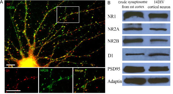
Background: Interactions between dopamine and glutamate in the prefrontal cortex are essential for cognitive functions such as working memory. Modulation of N-methyl-D-aspartic acid (NMDA) receptor functions by dopamine D1 receptor is believed to play a critical role in these functions. The aim of the work reported here is to explore the signaling pathway underlying D1 receptor-mediated trafficking of NMDA receptors in cultured rat prefrontal cortical neurons.

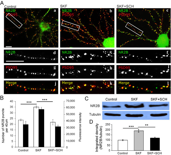
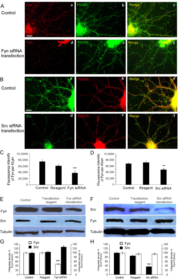
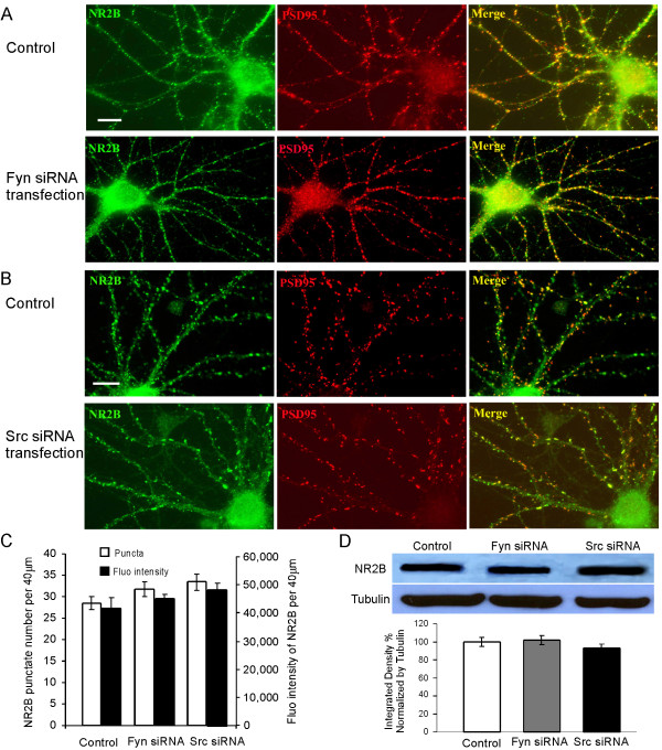
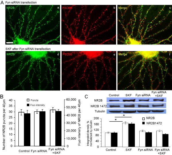
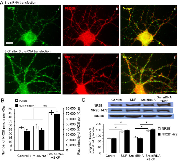
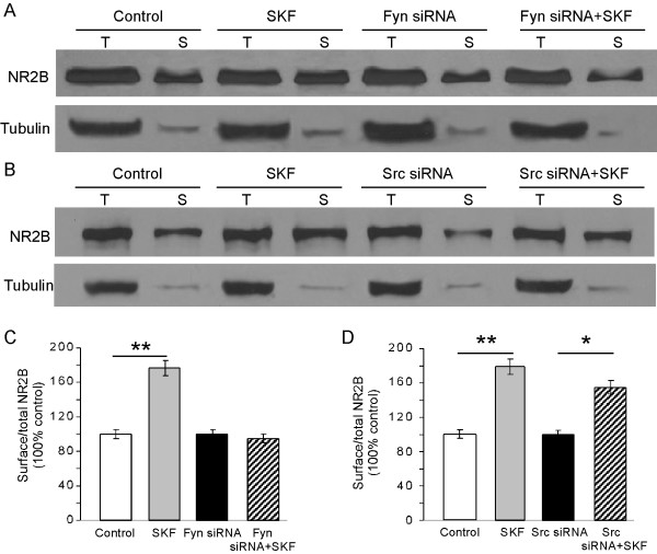
Results: Activation of D1 receptor by selective agonist SKF-81297 significantly increased the expression of NR2B subunits. This effect was completely blocked by small interfering RNA knockdown of Fyn, but not Src. Under control conditions, neither Fyn nor Src knockdown exhibited significant effect on basal NR2B expression. D1 stimulation significantly enhanced NR2B insertion into plasma membrane in cultured PFC neurons, a process obstructed by Fyn, but not Src, knockdown.

Conclusions: Dopamine D1 receptor-mediated increase of NMDA receptors is thus Fyn kinase dependent. Targeting this signaling pathway may be useful in treating drug addiction and schizophrenia.


Figures

Figure 1
Figure 1
Expression of D1 and NMDA receptors in cultured PFC neurons. (A) Localization of D1 receptor and NR2B in cultured PFC neurons. PFC neurons in dissociated culture at 14 DIV were labeled for endogenous D1 and NR2B receptors with double immunofluorescent staining. Lower panel, images at higher magnification showing the colocalization of D1 and NR2B on the dendritic shafts and spines. Scale bars = 10 μm. (B) Analysis by Western blotting shows the protein expression of D1 receptor and NMDA receptor subunits in neuronal lysates. Crude synaptosome (20 μg of protein) from adult rat cortex (lane 1) and 14 DIV cultured PFC neurons (lane 2) were subjected to sodium dodecyl sulfate-polyacrylamide gel electrophoresis (SDS-PAGE) and probed for NR1, NR2A, NR2B, D1, PSD95, and adaptin. Both adult rat cortex crude synptosome and cultured PFC neurons exhibited similar expressions of NMDA and D1 receptors, as well as synaptic protein, suggesting the validity of the experiments in primary cultured neurons.
Figure 2
Figure 2
D1 receptor agonist SKF-81297 enhances the expression and clustering of NR2B subunit. (A) PFC neurons at 16 DIV were treated with vehicle (a, d, g, j), SKF-81297 (10 μM; b, e, h, k), or SKF in the presence of SCH (15 μM; c, f, i, l), respectively, and double labeled for endogenous NR2B (green) and PSD95 (red). Panel j, k and l are merged from respective NR2B (green) and PSD-95(red) staining. Scale bars = 10 μm. (B) Quantification of total NR2B immunofluorescence staining. White bars: NR2B puncta number; black bars: NR2B fluorescence intensity. SKF-81297 significantly increased total NR2B puncta number and immunofluorescence intensity compared with control. Results are presented as mean number and fluorescence intensity of total NR2B puncta. Statistical analysis was performed using ANOVA followed by Tukey multiple comparison test (n = 20, ** p < 0.01, *** p < 0.001). Data represent mean ± SEM. (C) Proteins were isolated at 16 DIV from cultured high-density PFC neurons treated with DMSO, D1 receptor agonist SKF-81297, or SKF-81297 + SCH-23390. The proteins were resolved on SDS-PAGE and immunoblotted for NR2B and reprobed with tubulin. (D) Quantification of NR2B protein expression in PFC neurons. SKF-81297 significantly increased NR2B expression, which was completely blocked by pretreatment with SCH-23390 (n = 4, ** p < 0.01, *** p < 0.001). Integrated intensity was measured using Image J and control protein level of NR2B was set to 100% after being normalized to tubulin. These results indicate that D1 receptor stimulation does increase NR2B expression, which may eventually increase NMDA receptor trafficking to membrane surface.
Figure 3
Figure 3
Efficiency of Src and Fyn knockdown in cultured PFC neurons. (A) 11 DIV PFC neurons were transfected for 48 h with Fyn siRNA and stained with anti-Fyn and PSD95. Fyn, but not PSD95, decreased dramatically after siRNA transfection. Scale bar = 10 μm. (B) 11 DIV PFC neurons were transfected for 48 h with Src siRNA and stained with anti-Src and PSD95. Similarly, Src, but not PSD95, was significantly reduced in transfected neurons. Scale bar = 10 μm. (C and D) Quantifications of Fyn and Src immunofluorescence in dendrites suggest the effective knockdown of both Fyn and Src kinases in the cultured PFC neurons (n = 30, ** p < 0.01, *** p < 0.001). (E-H) Total protein levels of Fyn and Src were determined by Western blotting after Fyn and Src siRNA transfection in cultured PFC neurons. The blots were stripped and reprobed with antitubulin as loading control and the siRNA knockdown specificities were demonstrated by re-probing the Src and Fyn, respectively. Integrated intensity analysis showed significant decreases in both Fyn and Src proteins after knockdown (n = 3, ** p < 0.01). Control protein levels of Fyn and Src were set at 100% after being normalized to loading control tubulin.
Figure 4
Figure 4
Src and Fyn knockdowns have no effect on the overall expression or localization and clustering of NR2B subunit at basal conditions. (A) 11 DIV PFC neurons were transfected with Fyn siRNA for 48 h and stained for endogenous NR2B (green) and PSD95 (red). Scale bars = 10 μm. (B) 11 DIV PFC neurons were transfected with Src siRNA for 48 h and stained for endogenous NR2B (green) and PSD95 (red). Scale bars = 10 μm. (C) Quantification of total NR2B immunofluorescence staining showed that neither Fyn nor Src had a detectable effect on total NR2B puncta number or immunofluorescence intensity compared with the control group (p > 0.05). Data represent mean ± SEM. (D) Total protein levels of NR2B were determined by Western blotting after Fyn and Src siRNA transfection in cultured PFC neurons. The blots were stripped and reprobed with antitubulin as loading control. Control protein levels of NR2B were set at 100% after being normalized to loading control tubulin (p > 0.05).
Figure 5
Figure 5
D1 agonist induces no changes in NR2B expression after Fyn knockdown. Immunostaining revealed that neither NR2B nor PSD95 expression was changed by treatment with SKF-81297 (10 μM) in Fyn siRNA-transfected PFC neurons (d, e, f) compared with control (a, b, c). Scale bar = 10 μm. (B) Quantification of total NR2B immunofluorescence staining revealed that SKF-81297 (10 μM) had no clear effects on NR2B puncta number or fluorescence intensity in the dendrites of Fyn siRNA-transfected neurons compared with control group (p > 0.05). (C) Total protein levels of NR2B, which were determined by Western blotting after Fyn siRNA transfection followed by SKF treatment in cultured PFC neurons, were consistently unaltered by treatment with SKF-81297 (10 μM, p > 0.05) although SKF-81297 (10 μM) induced a significant increase in NR2B expression in the control condition (*p < 0.05). Similarly, in neurons with Fyn siRNA, NR2B Tyr1472 phosphorylation level was not changed by treatment with SKF-81297 (10 μM, p > 0.05). Integrated intensity analysis was performed using Image J software and the control protein level of NR2B or the Tyr1472 phosphorylation was set to 100% after being normalized by loading control tubulin.
Figure 6
Figure 6
Src knockdown has no effect on the D1 receptor-mediated localization and clustering of NR2B. (A) Immunostaining revealed that NR2B expression was significantly increased by SKF-81297 (10 μM) treatment in Src siRNA-treated PFC neurons, compared with the control group. Scale bar = 10 μm. (B) Quantification of total NR2B immunofluorescence staining revealed that SKF treatment increased total NR2B puncta number and fluorescence intensity in dendrites even after Src siRNA transfection, compared with control. Results are presented as the mean number of total NR2B puncta and mean intensity of fluorescence (**p < 0.01). Statistical analysis was performed by t test and the data represent mean ± SEM. (C) Similarly, total protein or NR2B Tyr1472 phosphorylation levels of NR2B determined by Western blotting after Src siRNA transfection remained to be significantly altered by SKF-81297 treatment in cultured PFC neurons (* p < 0.05). Control NR2B protein level or NR2B Tyr1472 phosphorylation level was set to 100% after being normalized by loading control tubulin.
Figure 7
Figure 7
Fyn, but not Src, affects the surface NR2B expression after D1 receptor stimulation. (A and B) Surface biotinylation of NMDA receptors in high-density PFC neurons at 14 DIV. PFC neurons at12 DIV were transfected with Fyn siRNA for 48 h. The PFC neurons at DIV 14 were treated with DMSO (0.1%, lanes 1 and 2); SKF-81297 (10 μM, lanes 3 and 4); Fyn (A) or Src (B) knockdown PFC neurons treated with DMSO (0.1%, lanes 5 and 6); or Fyn (A) or Src (B) knockdown neurons treated with SKF-81297 (10 μM, lanes 7 and 8). After surface receptor biotinylation, 20% of the lysis supernatant was used to detect the total (T) proteins. The remaining 80% of the supernatant was incubated with NeurAvidin Agarose beads and purified as surface (S) proteins. After SDS-PAGE the proteins were immunoblotted for NR2B and β-tubulin. (C and D) Quantification of surface NR2B expression. Surface NR2B was corrected by total NR2B to calculate the surface/total ratio and the control group was set to 100% for normalization. Fyn, but not Src, knockdown appeared to be effective in blocking the surface insertion of NR2B mediated by D1 stimulation (* p < 0.05, ** p < 0.01).

Similar articles

Cited by

References

    1. Goldman-Rakic PS, Castner SA, Svensson TH, Siever LJ, Williams GV. Targeting the dopamine D1 receptor in schizophrenia: insights for cognitive dysfunction. Psychopharmacology (Berl) 2004;174(1):3–16. doi: 10.1007/s00213-004-1793-y. - DOI - PubMed
    1. Castner SA, Williams GV. Tuning the engine of cognition: a focus on NMDA/D1 receptor interactions in prefrontal cortex. Brain Cogn. 2007;63(2):94–122. doi: 10.1016/j.bandc.2006.11.002. - DOI - PubMed
    1. Yang CR, Chen L. Targeting prefrontal cortical dopamine D1 and N-methyl-D-aspartate receptor interactions in schizophrenia treatment. Neuroscientist. 2005;11(5):452–70. doi: 10.1177/1073858405279692. - DOI - PubMed
    1. Missale C, Fiorentini C, Busi C, Collo G, Spano PF. The NMDA/D1 receptor complex as a new target in drug development. Curr Top Med Chem. 2006;6(8):801–8. doi: 10.2174/156802606777057562. - DOI - PubMed
    1. Cepeda C, Levine MS. Where do you think you are going? The NMDA-D1 receptor trap. Sci STKE. 2006;2006(333):pe20. doi: 10.1126/stke.3332006pe20. - DOI - PubMed

Publication types

MeSH terms

LinkOut - more resources