

Breast cancer stromal fibroblasts promote the generation of CD44+CD24- cells through SDF-1/CXCR4 interaction
- PMID: 20569497
- PMCID: PMC2911413
- DOI: 10.1186/1756-9966-29-80
Breast cancer stromal fibroblasts promote the generation of CD44+CD24- cells through SDF-1/CXCR4 interaction
Abstract
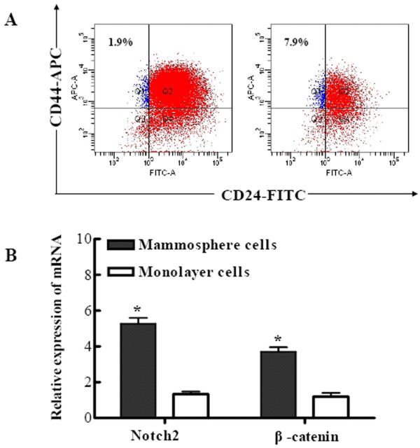
Background: Breast cancer stem cells (BCSCs) have been recently identified in breast carcinoma as CD44+CD24- cells, which exclusively retain tumorigenic activity and display stem cell-like properties. Using a mammosphere culture technique, MCF7 mammosphere cells are found to enrich breast cancer stem-like cells expressing CD44+CD24-. The stromal cells are mainly constituted by fibroblasts within a breast carcinoma, yet little is known of the contributions of the stromal cells to BCSCs.
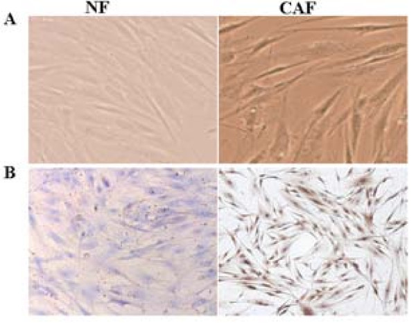
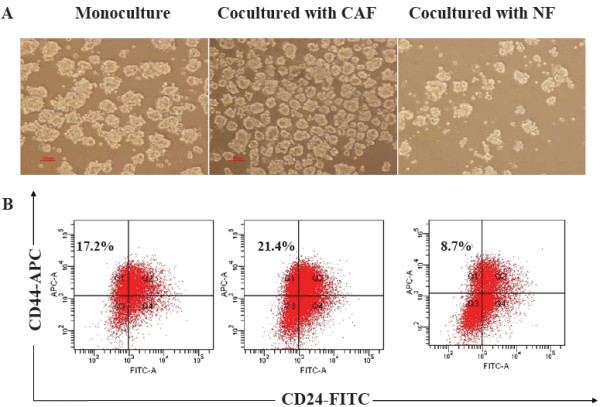
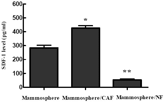
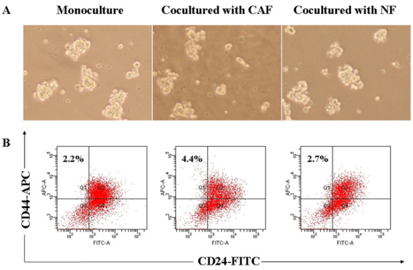
Methods: Carcinoma-associated fibroblasts (CAFs) and normal fibroblasts (NFs) were isolated and identified by immunohistochemistry. MCF7 mammosphere cells were co-cultured with different stromal fibroblasts by a transwell cocultured system. Flow cytometry was used to measure CD44 and CD24 expression status on MCF7. ELISA (enzyme-linked immunosorbent assay) was performed to investigate the production of stromal cell-derived factor 1 (SDF-1) in mammosphere cultures subject to various treatments. Mammosphere cells were injected with CAFs and NFs to examine the efficiency of tumorigenity in NOD/SCID mice.
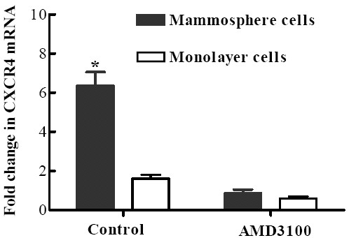
Results: CAFs derived from breast cancer patients were found to be positive for alpha-smooth muscle actin (alpha-SMA), exhibiting the traits of myofibroblasts. In addition, CAFs played a central role in promoting the proliferation of CD44+CD24- cells through their ability to secrete SDF-1, which may be mediated to SDF-1/CXCR4 signaling. Moreover, the tumorigenicity of mammosphere cells with CAFs significantly increased as compared to that of mammosphere cells alone or with NFs.
Conclusion: We for the first time investigated the effects of stromal fibroblasts on CD44+CD24- cells and our findings indicated that breast CAFs contribute to CD44+CD24- cell proliferation through the secretion of SDF-1, and which may be important target for therapeutic approaches.
Figures
Similar articles
-
SLUG/SNAI2 and tumor necrosis factor generate breast cells with CD44+/CD24- phenotype.BMC Cancer. 2010 Aug 6;10:411. doi: 10.1186/1471-2407-10-411. BMC Cancer. 2010. PMID: 20691079 Free PMC article.
-
Cancer-associated fibroblasts promote the progression of endometrial cancer via the SDF-1/CXCR4 axis.J Hematol Oncol. 2016 Feb 6;9:8. doi: 10.1186/s13045-015-0231-4. J Hematol Oncol. 2016. PMID: 26851944 Free PMC article.
-
Breast cancer-associated fibroblasts induce epithelial-to-mesenchymal transition in breast cancer cells.Endocr Relat Cancer. 2013 Jan 7;20(1):1-12. doi: 10.1530/ERC-12-0227. Print 2013 Feb. Endocr Relat Cancer. 2013. PMID: 23111755
-
Stromal fibroblasts in cancer: a novel tumor-promoting cell type.Cell Cycle. 2006 Aug;5(15):1597-601. doi: 10.4161/cc.5.15.3112. Epub 2006 Aug 1. Cell Cycle. 2006. PMID: 16880743 Review.
-
Prognosis assessment of CD44+/CD24- in breast cancer patients: a systematic review and meta-analysis.Arch Gynecol Obstet. 2022 Oct;306(4):1147-1160. doi: 10.1007/s00404-022-06402-w. Epub 2022 Apr 18. Arch Gynecol Obstet. 2022. PMID: 35435483
Cited by
-
CXCR4 activation maintains a stem cell population in tamoxifen-resistant breast cancer cells through AhR signalling.Br J Cancer. 2012 Jun 26;107(1):43-52. doi: 10.1038/bjc.2012.105. Epub 2012 May 29. Br J Cancer. 2012. PMID: 22644306 Free PMC article.
-
Baicalin promotes apoptosis and inhibits proliferation and migration of hypoxia-induced pulmonary artery smooth muscle cells by up-regulating A2a receptor via the SDF-1/CXCR4 signaling pathway.BMC Complement Altern Med. 2018 Dec 12;18(1):330. doi: 10.1186/s12906-018-2364-9. BMC Complement Altern Med. 2018. PMID: 30541517 Free PMC article.
-
Cancer-associated fibroblasts as therapeutic targets for cancer: advances, challenges, and future prospects.J Biomed Sci. 2025 Jan 9;32(1):7. doi: 10.1186/s12929-024-01099-2. J Biomed Sci. 2025. PMID: 39780187 Free PMC article. Review.
-
Exposure to ionizing radiation induced persistent gene expression changes in mouse mammary gland.Radiat Oncol. 2012 Dec 5;7:205. doi: 10.1186/1748-717X-7-205. Radiat Oncol. 2012. PMID: 23216862 Free PMC article.
-
Potential Strategies to Improve the Effectiveness of Drug Therapy by Changing Factors Related to Tumor Microenvironment.Front Cell Dev Biol. 2021 Aug 10;9:705280. doi: 10.3389/fcell.2021.705280. eCollection 2021. Front Cell Dev Biol. 2021. PMID: 34447750 Free PMC article. Review.
References
MeSH terms
Substances
LinkOut - more resources
Full Text Sources
Medical
Miscellaneous
