. 2010;12(3):R40.
doi: 10.1186/bcr2594. Epub 2010 Jun 22.

Proteomic and transcriptomic profiling reveals a link between the PI3K pathway and lower estrogen-receptor (ER) levels and activity in ER+ breast cancer

Affiliations

Proteomic and transcriptomic profiling reveals a link between the PI3K pathway and lower estrogen-receptor (ER) levels and activity in ER+ breast cancer

Chad J Creighton et al. Breast Cancer Res. 2010.

Abstract

Introduction: Accumulating evidence suggests that both levels and activity of the estrogen receptor (ER) and the progesterone receptor (PR) are dramatically influenced by growth-factor receptor (GFR) signaling pathways, and that this crosstalk is a major determinant of both breast cancer progression and response to therapy. The phosphatidylinositol 3-kinase (PI3K) pathway, a key mediator of GFR signaling, is one of the most altered pathways in breast cancer. We thus examined whether deregulated PI3K signaling in luminal ER+ breast tumors is associated with ER level and activity and intrinsic molecular subtype.

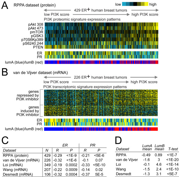
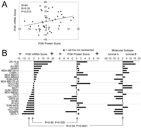
Methods: We defined two independent molecular signatures of the PI3K pathway: a proteomic (reverse-phase proteomic array) PI3K signature, based on protein measurement for PI3K signaling intermediates, and a PI3K transcriptional (mRNA) signature based on the set of genes either induced or repressed by PI3K inhibitors. By using these signatures, we scored each ER+ breast tumor represented in multiple independent expression-profiling datasets (four mRNA, n = 915; one protein, n = 429) for activation of the PI3K pathway. Effects of PI3K inhibitor BEZ-235 on ER expression and activity levels and cell growth were tested by quantitative real-time PCR and cell proliferation assays.

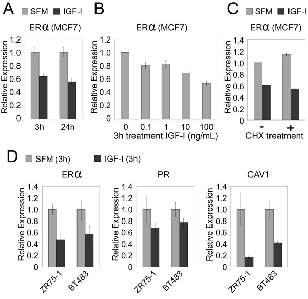
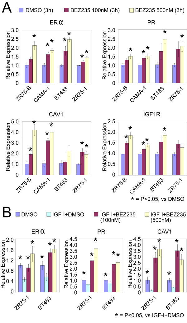
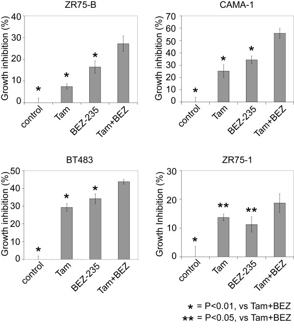
Results: Within ER+ tumors, ER levels were negatively correlated with the PI3K activation scores, both at the proteomic and transcriptional levels, in all datasets examined. PI3K signature scores were also higher in ER+ tumors and cell lines of the more aggressive luminal B molecular subtype versus those of the less aggressive luminal A subtype. Notably, BEZ-235 treatment in four different ER+ cell lines increased expression of ER and ER target genes including PR, and treatment with IGF-I (which signals via PI3K) decreased expression of ER and target genes, thus further establishing an inverse functional relation between ER and PI3K. BEZ-235 had an additional effect on tamoxifen in inhibiting the growth of a number of ER+ cell lines.

Conclusions: Our data suggest that luminal B tumors have hyperactive GFR/PI3K signaling associated with lower ER levels, which has been correlated with resistance to endocrine therapy. Targeting PI3K in these tumors might reverse loss of ER expression and signaling and restore hormonal sensitivity.


Figures

Figure 1
Figure 1
Proteomic and transcriptomic signatures of PI3K signaling are associated in ER+ breast tumors with lower ER and PR levels and the luminal B subtype. (a) Heat map of PI3K proteomic signature proteins in 429 ER+ human breast tumors, along with corresponding patterns for ER and PR (blue ER on the color scale meaning lower levels, although still present) and intrinsic molecular subtype association (luminal A versus luminal B). PI3K protein score is the sum of the phosphoprotein levels of Akt, mTOR, GSK3, S6K, and S6, minus the total levels of pathway-inhibitor PTEN. Tumors are ranked from low to high PI3K score, where a high PI3K score indicates high pathway activity. (b) PI3K transcriptomic (that is, mRNA) signature genes in 226 ER+ breast tumors (from van de Vijver et al.), along with ER and PR mRNA; tumors are ranked from low to high PI3K mRNA score. (c) Spearman's correlations between PI3K score and ER/PR in multiple expression-profiling datasets (four transcriptomic, one proteomic). For proteomic dataset, PI3K protein score and ER/PR protein levels were analyzed; for mRNA datasets, PI3K mRNA score and ER/PR mRNA. (d) In each of the five expression datasets, the average PI3K score in ER+ tumors of the luminal B subtype was compared with the average in ER+ tumors of the luminal A subtype by t test.
Figure 2
Figure 2
PI3K protein and mRNA activation scores correlate with each other and with the luminal B subtype in breast cancer cell lines. (a) Scatterplot of PI3K protein score versus PI3K mRNA score in 40 breast cancer cell lines. (b) Luminal breast cancer cell lines (as defined by Neve et al.) plotted alongside their PI3K mRNA score (left) and their PI3K protein score (middle), along with their correlation with a luminal B signature from Hoadley et al. (right). Note that some of the cell lines were not analyzed by proteomics and are indicated (*). Correlations (R values) by Spearman's.
Figure 3
Figure 3
PI3K-activator IGF-I rapidly downregulates mRNA levels of ER and ER target genes. (a) MCF-7 breast cancer cells were starved in SFM and then incubated with IGF-I (100 ng/ml, 8 nM) for 3 or 24 hours. RNA was isolated, and ER mRNA levels were measured with QRT-PCR (± SEM). (b) MCF-7 cells were incubated with an increasing dose of IGF-I, and ER mRNA levels were measured as in part (a). (c) ER levels in MCF-7 cells incubated with (+) or without (-) cycloheximide and stimulated with or without IGF-I (100 ng/ml) for 3 hours. (d) In two additional cell lines (ZR75-1 and BT483), expression levels of ER and ER target genes (PR and CAV1) after IGF-I treatment (100 ng/ml for 3 hours). All comparisons shown here between IGF-I (100 ng/ml) and control (± SD) were significant, with P ≤ 0.05.
Figure 4
Figure 4
Inhibition of PI3K signaling increases mRNA levels of ER and ER-inducible genes. (a) QRT-PCR results showing increased expression of ER and ER target genes (PR, CAV1, and IGF1R) in four different cell lines, after treatment with PI3K inhibitor BEZ-235 (100 nM and 500 nM for 3 hours). *Significant differences (P < 0.05; t test) between BEZ-235 and control (multiple comparisons expected to yield 32 × 0.05/23 = 7% false positives). Twelve of the 16 marker/cell-line combinations individually significant (P < 0.05) by ANOVA (P > 0.05: PR/ZR75-B, PR/ZR75-1, CAV1/BT483, IGF1R/ZR75-1; false-positive rate: 16 × 0.05/12 = 7%). (b) IGF-I-mediated downregulation of ER and ER target genes is restored by inhibition of PI3K. ZR75-1 and BT483 cells were preincubated for 30 minutes with or without BEZ-235 (100 and 500 nM) and then stimulated with or without IGF-I (100 ng/ml) for 3 hours. ER and ER targets gene mRNA levels were measured with QRT-PCR (data from DMSO and IGF-I + DMSO groups also are featured in Figure 3a). *Significant differences from corresponding IGF-I + DMSO group (P ≤ 0.05; t test). All marker/cell-line combinations were significant (P < 0.03) by ANOVA.
Figure 5
Figure 5
PI3K inhibitor BEZ-235 has an additional effect on tamoxifen (Tam) in ER+ cell lines. Growth-inhibition assays for luminal B-cell lines ZR75-B and CAMA-1 and luminal A cell lines BT483 and ZR75-1, treated for 4 days with Tam (100 nM), BEZ-235 (10 nM), or the combination of Tam + BEZ-235. Average growth inhibition normalized to control (regular medium) in each cell line (± SD). *Significant differences from Tam + BEZ-235 group (P < 0.05; t test). For each cell line, significant differences exist among the groups by ANOVA (P < 1E-5).

Comment in

Similar articles

Cited by

References

    1. Lin N, Winer E. Advances in adjuvant endocrine therapy for postmenopausal women. J Clin Oncol. 2008;26:798–805. doi: 10.1200/JCO.2007.15.0946. - DOI - PubMed
    1. Osborne C. Steroid hormone receptors in breast cancer management. Breast Cancer Res Treat. 1998;51:227–238. doi: 10.1023/A:1006132427948. - DOI - PubMed
    1. Cui X, Schiff R, Arpino G, Osborne CK, Lee AV. Biology of progesterone receptor loss in breast cancer and its implications for endocrine therapy. J Clin Oncol. 2005;23:7721–7735. doi: 10.1200/JCO.2005.09.004. - DOI - PubMed
    1. Conneely O, Jericevic B, Lydon J. Progesterone receptors in mammary gland development and tumorigenesis. J Mammary Gland Biol Neoplasia. 2003;8:205–214. doi: 10.1023/A:1025952924864. - DOI - PubMed
    1. Bardou V-J, Arpino G, Elledge RM, Osborne CK, Clark GM. Progesterone receptor status significantly improves outcome prediction over estrogen receptor status alone for adjuvant endocrine therapy in two large breast cancer databases. J Clin Oncol. 2003;21:1973–1979. doi: 10.1200/JCO.2003.09.099. - DOI - PubMed

Publication types

MeSH terms