. 2006 Mar 15;12(1):45-51.
doi: 10.1177/159101990601200109. Epub 2006 Jun 15.

Carotid-cavernous fistula in ehlers-danlos syndrome by pure transvenous approach

Affiliations

Carotid-cavernous fistula in ehlers-danlos syndrome by pure transvenous approach

O Van Overmeire et al. Interv Neuroradiol. .

Abstract

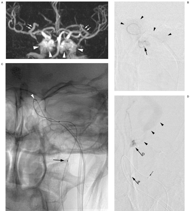
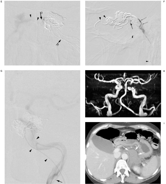
We describe a carotid-cavernous fistula (CCF) in a middle aged woman with Ehlers-Danlos syndrome (EDS) type IV, which manifested with a left-sided ophthalmoplegia. The CCF was diagnosed on magnetic resonance imaging. To prevent potential lethal arterial wall injury, the CCF was treated endovascularly under local anesthesia and exclusively by a transvenous approach. The fistula was successfully closed with Guglielmi Detachable Coils. Notwithstanding these precautionary measures, the patient suffered an intraperitoneal and a small retroperitoneal bleed during the procedure and died suddenly ten days after intervention in hemorrhagic shock. A review of recent literature focussing on the technique of transvenous approach and the catheterization risks of CCF in Ehlers-Danlos syndrome is presented.


Figures

Figure 1
Figure 1
A) Time-of-flight MR angiography revealing arterialization of both cavernous sinus (arrowheads) and the superior ophthalmic veins (arrows) as an indication of a CCF. Whether the CCF is unilateral left or right-sided or bilateral cannot be determined. B) DSA of the left internal carotid via the microcatheter (arrow) introduced through the inferior petrous sinus and positioned across the fistula hole. Contrast medium is vanishing into the cavernous sinus (arrowheads). No intracranial circulation is visualized. C) Digital image of the microcatheter stabilized with a microguidewire in the left internal carotid artery. Radiopaque marker (arrow) indicates tip of the microcatheter. Bend in the microcatheter indicating the fistula hole (arrowhead). D) DSA (frontal view) of the superior ophthalmic vein showing dilatation and flow reversal (arrowheads). Coiling starts at the outlet of the ophthalmic vein to prevent persistent flow and orbital hypertension in case occlusion of the CCF would be incomplete. Proximal marker (arrowP) and distal marker (or tip) of the coiling microcatheter (arrowD) are shown. Small arrows indicate the two markers on the microcatheter in the internal carotid artery. E) DSA of the left cavernous sinus showing high outflow to the contralateral sinus. The microcatheter is positioned adjacent to the delivered coils in the superior anterior part at the entry of the intermedial part (arrowD). Coiling will continue from this position. F) Control DSA of the cavernous sinus showing stagnation of contrast medium (small arrows). Coils are covering the fistula hole (arrow), which is landmarked by the bend in the microcatheter (small arrows) in the internal carotid artery. Coils in the outlet of the superior ophthalmic vein (arrowheads). G) Control DSA of the internal carotid artery via the microcatheter (arrowheads) showing intracranial circulation and confirming occlusion of the CCF, tip of the microcatheter (arrow), and petrous sinus track (two arrowheads). H) Time-of-flight MRI angiography after coiling only depicting arterial anatomy compatible with occlusion of the CCF. Short interruption of vessel continuity in the left carotid sinus is due to susceptibility artefacts of the coils. I) Contrast-enhanced CT scan of the abdomen shows retroperitoneal hemorrhage (white arrows). Blood around the spleen is also observed.
Figure 1
Figure 1
A) Time-of-flight MR angiography revealing arterialization of both cavernous sinus (arrowheads) and the superior ophthalmic veins (arrows) as an indication of a CCF. Whether the CCF is unilateral left or right-sided or bilateral cannot be determined. B) DSA of the left internal carotid via the microcatheter (arrow) introduced through the inferior petrous sinus and positioned across the fistula hole. Contrast medium is vanishing into the cavernous sinus (arrowheads). No intracranial circulation is visualized. C) Digital image of the microcatheter stabilized with a microguidewire in the left internal carotid artery. Radiopaque marker (arrow) indicates tip of the microcatheter. Bend in the microcatheter indicating the fistula hole (arrowhead). D) DSA (frontal view) of the superior ophthalmic vein showing dilatation and flow reversal (arrowheads). Coiling starts at the outlet of the ophthalmic vein to prevent persistent flow and orbital hypertension in case occlusion of the CCF would be incomplete. Proximal marker (arrowP) and distal marker (or tip) of the coiling microcatheter (arrowD) are shown. Small arrows indicate the two markers on the microcatheter in the internal carotid artery. E) DSA of the left cavernous sinus showing high outflow to the contralateral sinus. The microcatheter is positioned adjacent to the delivered coils in the superior anterior part at the entry of the intermedial part (arrowD). Coiling will continue from this position. F) Control DSA of the cavernous sinus showing stagnation of contrast medium (small arrows). Coils are covering the fistula hole (arrow), which is landmarked by the bend in the microcatheter (small arrows) in the internal carotid artery. Coils in the outlet of the superior ophthalmic vein (arrowheads). G) Control DSA of the internal carotid artery via the microcatheter (arrowheads) showing intracranial circulation and confirming occlusion of the CCF, tip of the microcatheter (arrow), and petrous sinus track (two arrowheads). H) Time-of-flight MRI angiography after coiling only depicting arterial anatomy compatible with occlusion of the CCF. Short interruption of vessel continuity in the left carotid sinus is due to susceptibility artefacts of the coils. I) Contrast-enhanced CT scan of the abdomen shows retroperitoneal hemorrhage (white arrows). Blood around the spleen is also observed.

References

    1. Beighton P, De Paepe A, et al. Ehlers-Danlos syndromes: revised nosology, Villefranche, 1997. Ehlers-Danlos National Foundation (USA) and Ehlers-Danlos Support Group (UK) Am J Med Genet. 1998;77:31–37. - PubMed
    1. De Paepe A, Malfait F. Bleeding and bruising in patients with Ehlers-Danlos syndrome and other collagen vascular disorders. Br J Haematol. 2004;127:491–500. - PubMed
    1. Citron SJ, Wallace RC, et al. Quality improvement guidelines for adult diagnostic neuroangiography: cooperative study between ASITN, ASNR, and SIR. J Vasc Interv Radiol. 2003;14:S257–262. - PubMed
    1. Schievink WI, Piepgras DG, et al. Spontaneous carotid-cavernous fistulae in Ehlers-Danlos syndrome Type IV. Case report. J Neurosurg. 1991;74:991–998. - PubMed
    1. Lach B, Nair SG, et al. Spontaneous carotid-cavernous fistula and multiple arterial dissections in type IV Ehlers-Danlos syndrome. Case report. J Neurosurg. 1987;66:462–467. - PubMed

LinkOut - more resources