. 2006 Sep 15;12(3):189-202.
doi: 10.1177/159101990601200301. Epub 2006 Dec 13.

Neurovascular radiosurgery

Affiliations

Neurovascular radiosurgery

M Söderman et al. Interv Neuroradiol. .

Abstract

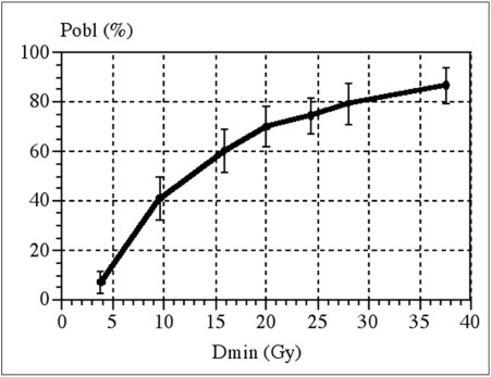
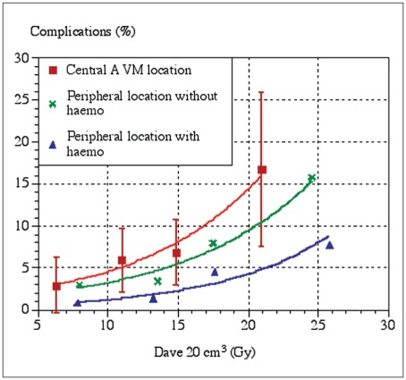
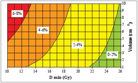
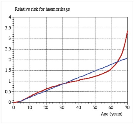
This article focuses on the treatment of neurovascular diseases, in particular brain arteriovenous malformations (BAVMs), with radiosurgery. The target group for this review is physicians who manage patients with neurovascular diseases, but are not actively engaged in radiosurgery. Radiosurgery for BAVMs is an established treatment with clearly defined risks and benefits. The efficacy of radiosurgery for dural arteriovenous shunts (DAVSs) is probably similar but the treatment has not yet gained the same acceptance. Radiosurgical treatment of cavernomas (cavernous hemangiomas) remains controversial. Well founded predictive models for BAVM radiosurgery show: * The probability of obliteration depends on the dose of radiation given to the periphery of the BAVM. * The risk of adverse radiation effects depends on the total dose of radiation, i.e. the amount of energy imparted into the tissue. The risk is greater in centrally located lesions. The risk of damage to brainstem nucleii and cranial nerves must be added to the risk predicted from current outcome models. * The risk of hemorrhage during the time span before obliteration depends on the BAVM volume, the dose of radiation to the periphery of the lesion and the age of the patient. Central location is a probably also a risk factor.


Figures

Figure 1
Figure 1
A) Vertebral angiography, posteroanterior view. A poorly defined BAVM is present in the quadrigeminal cistern, draining into the precentral vein. B) Vertebral angiography, lateral view. The same BAVM is visible. C) Biplane angiography in stereotactic frame. The final dose plan is projected onto the angiographic images. The 20 Gy isodose is depicted in yellow and and the 10 Gy isodose in green. D) The same dose plan has been projected onto the stereotactic MRI. The red and blue lines represent the projections from the stereotactic angiography. Note how the 20 Gy isodose line (yellow) has been shaped to avoid excessive irradiation to the quadrigeminal plate. The dose fall off is 10 Gy within 5 mm (green line). The left superior quadrigeminal body was damaged by the initial hemorrhage and the patient had permanent diplopia. Therefore was accepted a slightly higher than usual radiation dose to the left side of the quadrigeminal plate. The BAVM nidus was not discernible on the MRI, performed without and with Gadolinium and including MR angiography. At angiography two years after the treatment the BAVM had obliterated. There was no complication to the treatment and no rehemorrhage.
Figure 2
Figure 2
Rolandic BAVM, previously embolized at another institution with occlusion of the Rolandic artery. The BAVM was supplied by an extensive pial arterial collateral system. A) Angiography with injection into the left internal carotid artery. Lateral view in the early phase of contrast media passage, just as the vein becomes visible. Two distinct shunting zones are evident (arrows) which were separately delineated before GKRS. B) Later phase of the angiogram, when the vein is filled with contrast medium. It is no longer possible to distinguish the shunts. The two separate target delineations are shown on this image. C) Posteroanterior view in a late phase of the angiogram with the two separate target delineations. Treatment was with 25Gy to the 50% isodose. D) After two years the BAVM was obliterated. There is no remaining arteriovenous shunt. Note that the pial arterial collateral network has regressed almost totally.
Figure 3
Figure 3
Two examples of 100% coverage of an ellipsoid volume. In the first case there is good conformity to the volume (grey ellipsoid) that has been covered with multiple lesions (dotted circles). In the second case the volume has been covered with only one single spherical lesion (black circle with blue background) as is common with less sophisticated or old LINACs. Both treatments will be with the same nominal minimum dose to the periphery of the target, but the amount of tissue irradiated and the dose distribution will differ significantly between the two "identical" treatments.
Figure 4
Figure 4
The relation between minimum dose to the periphery of the BAVM and likelihood of obliteration over a 2-year period. Reproduced with permission from Ref. (80).
Figure 5
Figure 5
MRI of a left frontal BAVM treated with a dose of 25Gy to the periphery in the GK. A) T2-weighted image at the time of the treatment. B) T2-weighted image eight months after the treatment. The patient developed seizures. C) T2-weighted image 12 months after the treatment. The patient was on steroids and anticonvulsive medication. D) T2-weighted image 24 months after the treatment. The patient was no longer on steroids or anticonvulsive drugs. E) T1-weighted image without Gadolinium 24 months after the treatment. F) T1-weighted image with Gadolinium 24 months after the treatment. Note the contrast enhancement at the position of the previously treated BAVM. The malformation was proven by angiography to be obliterated.
Figure 6
Figure 6
The risk of radiation-induced complications as a function of the average dose in a 20cm3 volume containing the target and the surrounding tissues. Due to the small number of complications the confidence intervals are large. Reproduced with permission from Ref. (79).
Figure 7
Figure 7
T1-weighted image with Gadolinium. Typical late post radiosurgery pseudocyst with an enhancing, more solid portion in the adjacent brain. The BAVM had been proven by angiography to be cured. The enhancing scar tissue was extirpated and the cyst was marsupialized. The patient recovered uneventfully.
Figure 8
Figure 8
The relation between the risk of hemorrhage during the two year latency period, BAVM volume and minimum radiation dose. Reproduced, with permission, from Ref. (81). The risk is also age-related. Therefore the calculated risk in the graph should be multiplied by the age-related relative risk factor from Fig. 9.
Figure 9
Figure 9
The age-related risk factor for BAVM hemorrhage. Reproduced with permission from Ref. (81).

References

    1. Hillman J. Population-based analysis of arteriovenous malformation treatment. J Neurosurg. 2001;95(4):633–637. - PubMed
    1. Jellinger K. Vascular malformations of the central nervous system: a morphological overview. Neurosurg Rev. 1986;9(3):177–216. - PubMed
    1. Karhunen PJ, Penttila A, Erkinjuntti T. Arteriovenous malformation of the brain: imaging by postmortem angiography. Forensic Sci Int. 1990;48(1):9–19. - PubMed
    1. Wikholm G, Lundqvist C, Svendsen P. Embolization of cerebral arteriovenous malformations: Part I-Technique, morphology, and complications. Neurosurgery. 1996;39(3):448–457. discussion 457-459. - PubMed
    1. Ondra SL, Troupp H, et al. The natural history of symptomatic arteriovenous malformations of the brain: a 24-year follow up assessment. Journal of Neurosurgery. 1990;73:387–391. - PubMed

LinkOut - more resources