

Effects of stenting the parent artery on aneurysm filling and gene expression of various potential factors involved in healing of experimental aneurysms
- PMID: 20569585
- PMCID: PMC3354598
- DOI: 10.1177/159101990601200401
Effects of stenting the parent artery on aneurysm filling and gene expression of various potential factors involved in healing of experimental aneurysms
Abstract
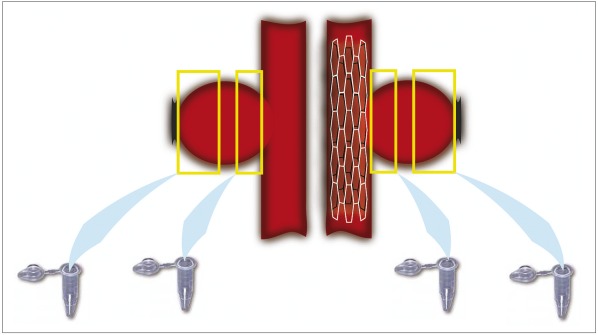
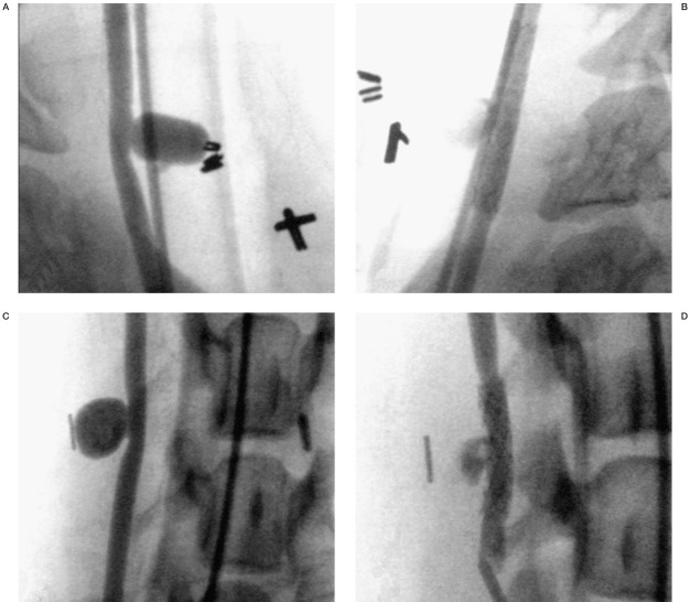
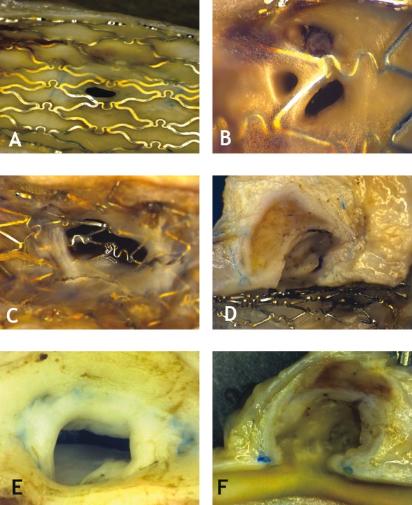
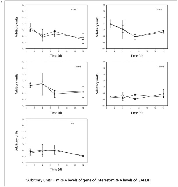
Intracranial stents are increasingly used in the endovascular treatment of aneurysms, but very little is known regarding their effect on the cellular and molecular evolution of aneurysms. Bilateral venous pouch lateral wall carotid aneurysms were created in 20 dogs. All dogs then underwent angiography and balloon-expandable stenting of one aneurysm four to six weeks later. Fifteen dogs underwent aneurysm harvesting at one day (n=3), four days (n=4), seven days (n=3), and 14 days (n=5) for mRNA expression analysis, using axial sections taken from the aneurysm neck and fundus for RTPCR amplification of four cytokines or growth factors: TNF-a, TGF-b1, MCP-1, and PDGFBB; two adhesion molecules: VCAM-1 and PECAM-1; five matrix modifying agents; MMP- 2, 9, TIMPs 1, 3, 4, and two cellular markers: CD34 and a-SMA. Five other dogs, sacrificed at 12 weeks, were examined for extent of filling of the aneurysm neck with organized tissue and for neointima formation at the aneurysm ostium. Angiography was performed prior to sacrifice in all animals, and compared with initial studies. Eleven out of 20 stented aneurysms showed a favorable angiographic evolution, while none of the 20 nonstented aneurysms improved (p=0.001). Pathology showed partially occluded aneurysms, with neointima formation around the stent struts.Observed trends in mRNA expression, that stenting increased expression of genes involved in organization and neointima formation, agreed with experimental hypotheses, but differences between stented and non-stented aneurysms did not reach statistical significance. Parent vessel stenting was associated with angiographic improvement of aneurysm appearance. Modifications in mRNA expression patterns following stenting deserve further study to better establish potential molecular targets to promote aneurysm healing.
Figures

Similar articles
-
The effects of stenting and endothelial denudation on aneurysm and branch occlusion in experimental aneurysm models.J Vasc Surg. 2007 Jun;45(6):1228-35. doi: 10.1016/j.jvs.2007.02.060. J Vasc Surg. 2007. PMID: 17543687
-
Endovascular treatment of aneurysms: gene expression of neointimal cells recruited on the embolic agent and evolution with recurrence in an experimental model.J Vasc Interv Radiol. 2005 Oct;16(10):1355-63. doi: 10.1097/01.RVI.0000171693.68581.96. J Vasc Interv Radiol. 2005. PMID: 16221907
-
Healing mechanisms in experimental aneurysms. I. Vascular smooth muscle cells and neointima formation.J Neuroradiol. 1999 Mar;26(1):7-20. J Neuroradiol. 1999. PMID: 10363438
-
Pathology of Stented Common Carotid Aneurysm in Dogs. Comparison between Stenting and Stent-Assisted Coiling.Interv Neuroradiol. 2005 Dec 20;11(4):333-40. doi: 10.1177/159101990501100405. Epub 2006 Feb 10. Interv Neuroradiol. 2005. PMID: 20584445 Free PMC article.
-
Complex intracranial aneurysms: combined operative and endovascular approaches.Neurosurgery. 1998 Dec;43(6):1304-12; discussion 1312-3. doi: 10.1097/00006123-199812000-00020. Neurosurgery. 1998. PMID: 9848843 Review.
Cited by
-
Various techniques of stent-assisted coil embolization of wide-necked or fusiform middle cerebral artery aneurysms : initial and mid-term results.J Korean Neurosurg Soc. 2013 May;53(5):274-80. doi: 10.3340/jkns.2013.53.5.274. Epub 2013 May 31. J Korean Neurosurg Soc. 2013. PMID: 23908700 Free PMC article.
-
Thrombosis heralding aneurysmal rupture: an exploration of potential mechanisms in a novel giant swine aneurysm model.AJNR Am J Neuroradiol. 2013 Feb;34(2):346-53. doi: 10.3174/ajnr.A3407. Epub 2012 Nov 15. AJNR Am J Neuroradiol. 2013. PMID: 23153870 Free PMC article.
-
Stent-assisted coiling of bifurcation aneurysms may improve endovascular treatment: a critical evaluation in an experimental model.AJNR Am J Neuroradiol. 2013 Mar;34(3):570-6. doi: 10.3174/ajnr.A3231. Epub 2012 Aug 16. AJNR Am J Neuroradiol. 2013. PMID: 22899786 Free PMC article. Clinical Trial.
-
Impact of the Transforming Growth Factor β (TGF-β) on Brain Aneurysm Formation and Development: A Literature Review.Cell Mol Neurobiol. 2025 May 20;45(1):46. doi: 10.1007/s10571-025-01572-y. Cell Mol Neurobiol. 2025. PMID: 40392340 Free PMC article. Review.
-
Comparison of 2-year angiographic outcomes of stent- and nonstent-assisted coil embolization in unruptured aneurysms with an unfavorable configuration for coiling.AJNR Am J Neuroradiol. 2011 Oct;32(9):1707-10. doi: 10.3174/ajnr.A2592. Epub 2011 Aug 18. AJNR Am J Neuroradiol. 2011. PMID: 21852378 Free PMC article.
References
-
- Molyneux A, et al. International Subarachnoid Aneurysm Trial (ISAT) of neurosurgical clipping versus endovascular coiling in 2143 patients with ruptured intracranial aneurysms: a randomised trial. Lancet, 2002;360(9342):1267–1274. - PubMed
-
- Raymond J, et al. Long-term angiographic recurrences after selective endovascular treatment of aneurysms with detachable coils. Stroke, 2003;34(6):1398–1403. - PubMed
-
- Lanzino G, et al. Emerging concepts in the treatment of intracranial aneurysms: stents, coated coils, and liquid embolic agents. Neurosurgery. 2005;57(3):449–459. discussion 449-459. - PubMed
LinkOut - more resources
Full Text Sources
Miscellaneous
