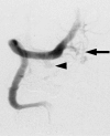
Transarterial embolization of a cervical dural arteriovenous fistula. Presenting with subarachnoid hemorrhage
- PMID: 20569588
- PMCID: PMC3354601
- DOI: 10.1177/159101990601200404
Transarterial embolization of a cervical dural arteriovenous fistula. Presenting with subarachnoid hemorrhage
Abstract
We describe a case of a 75-year-old man who presented with acute onset of headache and subarachnoid hemorrhage and initial cerebral angiography was deemed "negative". In retrospect, a faint contrast collection was present adjacent to the right vertebral artery at the C1 level suspicious for a small dural arteriovenous fistula (dAVF). Follow-up angiography with selective microcatheter injections of the right vertebral artery and C1 radicular artery confirmed a complex dAVF with characteristically specific venous drainage patterns associated with a subarachnoid hemorrhage presentation. Subsequently, the cervical dAVF was treated with superselective glue embolization resulting in complete occlusion. Cervical dAVFs are extremely rare vascular causes of subarachnoid hemorrhage. Both diagnostic angiography and endovascular treatment of these lesions can be challenging, especially in an emergent setting, requiring selective evaluation of bilateral vertebral arteries and careful attention to their cervical segments. Although only a single prior case of a cervical dAVF presenting with subarachnoid hemorrhage has been successfully treated with embolization, modern selective transarterial techniques may allow easier detection and treatment of subtle pathologic arteriovenous connections.
Figures


References
-
- Kendall BE, Logue V. Spinal epidural angiomatous malformations draining into intrathecal veins. Neuroradiology. 1977;13(4):181–189. - PubMed
-
- Symon L, Kuyama H, Kendall B. Dural arteriovenous malformations of the spine. Clinical features and surgical results in 55 cases. J Neurosurg. 1984;60(2):238–247. - PubMed
-
- Kinouchi H, Mizoi K, et al. Dural arteriovenous shunts at the craniocervical junction. J Neurosurg. 1998;89(5):755–761. - PubMed
-
- Reinges MH, Thron A, et al. Dural arteriovenous fistulae at the foramen magnum. J Neurol. 2001;248(3):197–203. - PubMed
LinkOut - more resources
Full Text Sources

