

doi: 10.1177/159101990601200412.
Epub 2007 Jan 19.
New Development of a Dural Arteriovenous Fistula (AVF) of the Superior Sagittal Sinus after Transvenous Embolization of a Left Sigmoid Sinus Dural AVF. Case Report and Review of the Literature
Affiliations
- PMID: 20569596
- PMCID: PMC3354609
- DOI: 10.1177/159101990601200412
Item in Clipboard
New Development of a Dural Arteriovenous Fistula (AVF) of the Superior Sagittal Sinus after Transvenous Embolization of a Left Sigmoid Sinus Dural AVF. Case Report and Review of the Literature
Interv Neuroradiol.
.
Abstract
Transvenous occlusion of an affected sinus has become a standardized curative treatment for dural sinus arteriovenous fistula. A 57-yearold man with a left sigmoid sinus isolated dural AVF was successfully treated with tansarterial followed by transvenous embolization. Followup angiography one year and two months thereafter showed complete disappearance of the dural AVF. However, one year later, superior parasagittal sinus dural arteriovenous fistula had newly developed, for which the etiology and a careful point for follow-up are here discussed.
Figures
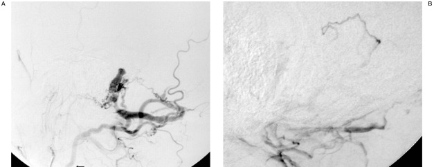
Left external carotid angiogram (lateral view) A) Early phase and B) late phase) showing an isolated dural AVF in the sigmoid sinus, draining into cortical vein and left external jugular vein via the deep cervical vein. The proximal part of the left jugular vein is tortuous and narrow (arrow).
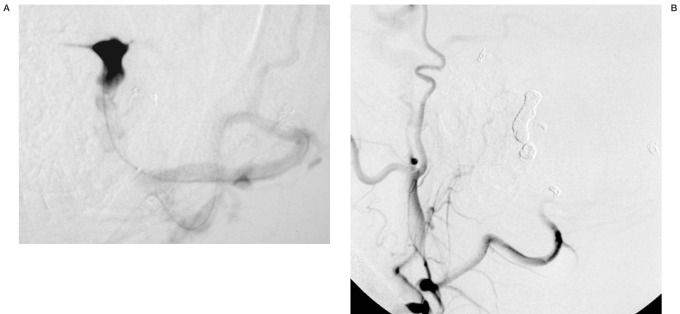
A) Venogram (lateral view) of the isolated sinus achieved by direct puncture of the left external jugular vein beyond the narrowing part. B) A left external carotid angiogram after transvenous embolization.
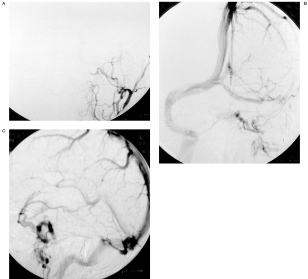
Follow-up left external carotid angiograms (anterior-posterior view) A) artertial phase, B) venous phase) (lateral view) C) venous phase) one year and two months after the TVE showing persistent disappearance of the dural AVF.
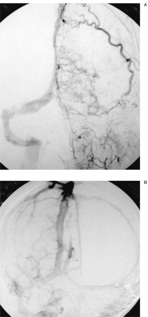
A:) A left external carotid angiogram showing persistent disappearance of the previously treated dural AVF at the left sigmoid sinus and a newly developed dural AVF at the superior sagittal sinus supplied by the left superficial temporal artery. B) A right internal carotid angiogram (venous phase), showing left transverse sinus and superior sagittal sinus.
References
-
- Houser OW, Cambell JK, et al. Arteriovenous malformation affecting the transverse dural venous sinus: an aquired lesion. Mayo Clin Proc. 1979;54:651–661. - PubMed
LinkOut - more resources
Full Text Sources
