

doi: 10.1177/15910199060120S123.
Epub 2006 Jun 15.
Treatment of vertebro-basilar dissecting aneurysms using intravascular stents
Affiliations
- PMID: 20569619
- PMCID: PMC3387941
- DOI: 10.1177/15910199060120S123
Item in Clipboard
Treatment of vertebro-basilar dissecting aneurysms using intravascular stents
Interv Neuroradiol.
.
Abstract
Endovascular surgery is an established primary therapeutic modality for dissecting aneurysms at vertebro-basilar arteries. Intravascular stents can be used to treat the dissecting aneurysms for which simple obliteration procedures cannot be used. In such cases, stent implantation alone or a combination of stents and coils need to be selected properly by taking into consideration the site and shape of dissections. In this report, three patterns of stent application are described and their method of selection is discussed.
Figures
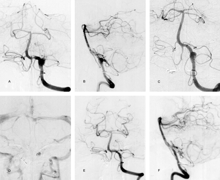
Case 1: Left VAG before treatment (A,B), after the second stent placement (C,D), and 3.5 months after the initial treatment (E,F). The dissecting aneurysm involved branching of the left PICA. Stagnation of the blood flow is shown soon after the second stent implantation (D). Complete obliteration of the aneurysm while preserving the left VA and PICA was obtained 2.5 months after placement of the second stent.
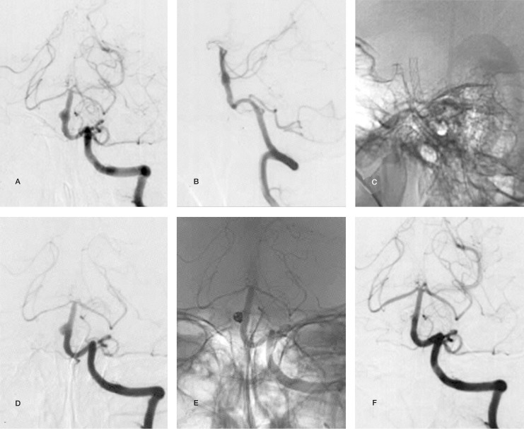
Case 2: Left VAG before treatment (A,B), after stent implantation (C), and four weeks after stent implantation (D) and a plain roentgen graph after addition of coils (E). The aneurysm was obliterated six months after the initial treatment (F).
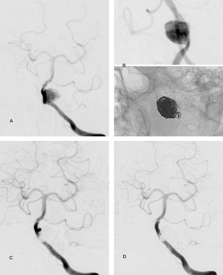
Case 3: A large dissecting aneurysm at the left VA was treated with a stent and coils (A,B). A small remnant neck was shown after coil embolizaion (C), and persisted for more than six months. Follow-up examination at 12 months showed complete obliteration (D).
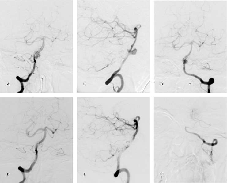
Case 4: Right (A,B) and left (C) VAG before treatment. Right VAG (D,E) immediately after the implantation of a stent from the BA to right VA and coil embolization of the aneurysm and left VA (F).

Three patterns of stent implantation used in this report: A) single stent or double stent implantation, B,C) combinations of a stent and coils. Broken lines show occlusive portions and gray zones show area that should be thrombosed.
Similar articles
-
[Combined endovascular stent implantation and coil embolization for the treatment of a vertebro-basilar fusiform aneurysm: technical case report].No Shinkei Geka. 2000 Sep;28(9):811-6. No Shinkei Geka. 2000. PMID: 11025882 Review. Japanese.
-
[Endovascular management and classification of the dissecting aneurysms of the vertebral artery].Zhonghua Yi Xue Za Zhi. 2017 Jun 20;97(23):1773-1777. doi: 10.3760/cma.j.issn.0376-2491.2017.23.004. Zhonghua Yi Xue Za Zhi. 2017. PMID: 28647997 Chinese.
-
Combined endovascular treatment of dissecting vertebral artery aneurysms by using stents and coils.J Neurosurg. 2001 Mar;94(3):427-32. doi: 10.3171/jns.2001.94.3.0427. J Neurosurg. 2001. PMID: 11235947 Clinical Trial.
-
Intravascular stent and endovascular coil placement for a ruptured fusiform aneurysm of the basilar artery. Case report and review of the literature.J Neurosurg. 1997 Dec;87(6):944-9. doi: 10.3171/jns.1997.87.6.0944. J Neurosurg. 1997. PMID: 9384409 Review.
-
Efficacy and current limitations of intravascular stents for intracranial internal carotid, vertebral, and basilar artery aneurysms.J Neurosurg. 1999 Oct;91(4):538-46. doi: 10.3171/jns.1999.91.4.0538. J Neurosurg. 1999. PMID: 10507372
Cited by
-
Management of posterior fossa dissecting aneurysms.Interv Neuroradiol. 2008 Nov 11;14 Suppl 2(Suppl 2):65-74. doi: 10.1177/15910199080140s212. Epub 2009 Jan 2. Interv Neuroradiol. 2008. PMID: 20557803 Free PMC article.
References
-
- Anxionnat R, Neto JFM, et al. Treatment of haemorrhagic dissections. Neurosurgery. 2001;53:289–301. - PubMed
-
- Han PP, Albuquerque FC, et al. Percutaneous intracranial stent placement for aneurysms. J Neurosurg. 2003;99:2330. - PubMed
-
- Higashida RT, Smith W, et al. Intravascular stent and endovascular coil placement for a ruptured fusiform aneurysm of the basilar artery. J Neurosurg. 1997;87:944–949. - PubMed
LinkOut - more resources
Full Text Sources
