

Orbitofrontal volume reductions during emotion recognition in patients with major depression
- PMID: 20569645
- PMCID: PMC2928284
- DOI: 10.1503/jpn.090076
Orbitofrontal volume reductions during emotion recognition in patients with major depression
Abstract
Background: Major depressive disorder is associated with both structural and functional alterations in the emotion regulation network of the central nervous system. The relation between structural and functional changes is largely unknown. Therefore, we sought to determine the relation between structural differences and functional alterations during the recognition of emotional facial expressions.
Methods: We examined 13 medication-free patients with major depression and 15 healthy controls by use of structural T1-weighted high-resolution magnetic resonance imaging (MRI) and functional MRI during 1 session. We set the statistical threshold for the analysis of imaging data to p < 0.001 (uncorrected).
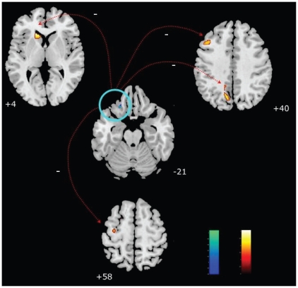
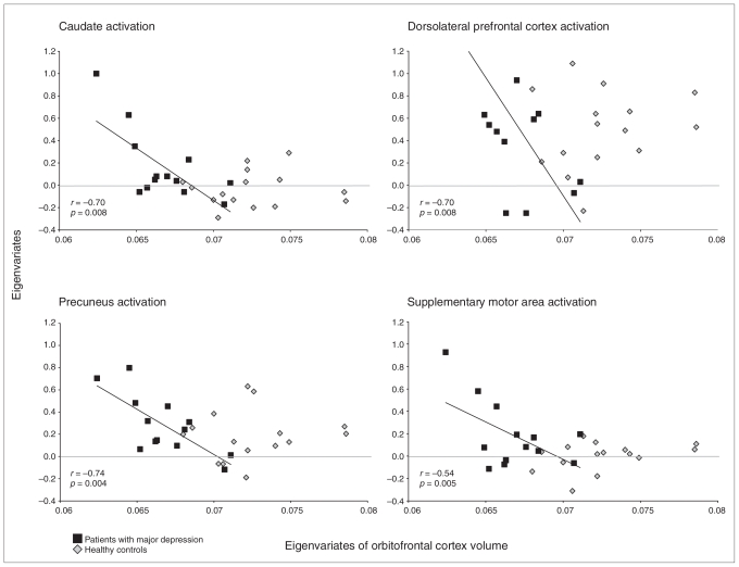
Results: As shown by voxel-based morphometry, depressed patients had reductions in orbitofrontal cortex volume and increases in cerebellar volume. Additionally, depressed patients showed increased activity during emotion recognition in the middle frontal cortex, caudate nucleus, precuneus and lingual gyrus. Within this cerebral network, the orbitofrontal volumes were negatively correlated in depressed patients but not in healthy controls with changes in blood oxygen level-dependent signal in the middle frontal gyrus, caudate nucleus, precuneus and supplementary motor area.
Limitations: Our results are limited by the relatively small sample size.
Conclusions: This combined functional and structural MRI study provides evidence that the orbitofrontal cortex is a key area in major depression and that structural changes result in functional alterations within the emotional circuit. Whether these alterations in the orbitofrontal cortex are also related to persistent emotional dysfunction in remitted mental states and, therefore, are related to the risk of depression needs further exploration.
Figures

References
-
- Gotlib IH, Krasnoperova E, Yue DN, et al. Attentional biases for negative interpersonal stimuli in clinical depression. J Abnorm Psychol. 2004;113:121–35. - PubMed
-
- Persad SM, Polivy J. Differences between depressed and nondepressed individuals in the recognition of and response to facial emotional cues. J Abnorm Psychol. 1993;102:358–68. - PubMed
-
- Frodl T, Moller HJ, Meisenzahl E. Neuroimaging genetics: New perspectives in research on major depression? Acta Psychiatr Scand. 2008;118:363–72. - PubMed
-
- Coffey CE, Wilkinson WE, Weiner RD, et al. Quantitative cerebral anatomy in depression. A controlled magnetic resonance imaging study. Arch Gen Psychiatry. 1993;50:7–16. - PubMed
-
- Lacerda ALT, Keshavan MS, Hardan AY, et al. Anatomic evaluation of the orbitofrontal cortex in major depressive disorder. Biol Psychiatry. 2004;55:353–8. - PubMed
Publication types
MeSH terms
LinkOut - more resources
Full Text Sources
Research Materials