. 2010 Jun 4;6(6):535-46.
doi: 10.1016/j.stem.2010.05.003.

A murine ESC-like state facilitates transgenesis and homologous recombination in human pluripotent stem cells

Affiliations

A murine ESC-like state facilitates transgenesis and homologous recombination in human pluripotent stem cells

Christa Buecker et al. Cell Stem Cell. .

Abstract

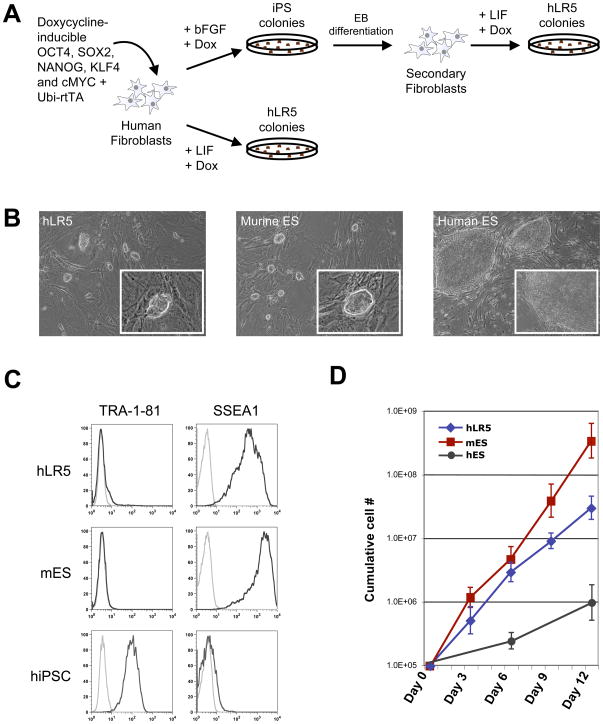
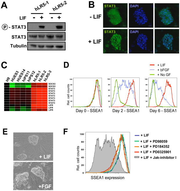
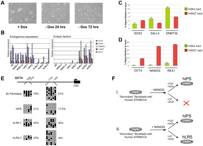
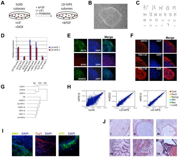
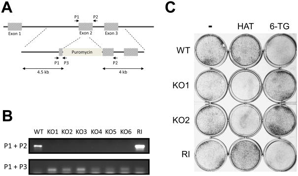
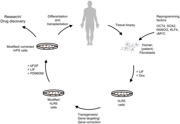
Murine pluripotent stem cells can exist in two functionally distinct states, LIF-dependent embryonic stem cells (ESCs) and bFGF-dependent epiblast stem cells (EpiSCs). However, human pluripotent cells so far seemed to assume only an epiblast-like state. Here we demonstrate that human iPSC reprogramming in the presence of LIF yields human stem cells that display morphological, molecular, and functional properties of murine ESCs. We termed these hLR5 iPSCs because they require the expression of five ectopic reprogramming factors, Oct4, Sox2, Klf4, cMyc, and Nanog, to maintain this more naive state. The cells are "metastable" and upon ectopic factor withdrawal they revert to standard human iPSCs. Finally, we demonstrate that the hLR5 state facilitates gene targeting, and as such provides a powerful tool for the generation of recombinant human pluripotent stem cell lines.


Figures

Figure 1
Figure 1. A metastable human iPS state with murine ES cell properties
(A) Schematic representation of the used strategy.. Doxycycline-inducible lentiviral vectors were added either as individual vectors (Maherali et al., 2008) or the polycystronic human STEMCCA virus (Sommer et al., 2009) and inducible NANOG as indicated in the text (B) Colony morphology of hLR5 cells (left panel), murine ES cells (middle panel) and human ES cells. (C) FACS analysis of cell surface marker expression on hLR5 cells, murine ES cells and human iPS cells. Black lines: Cell surface marker using the indicated primary antibody. Grey line: no primary antibody control. (D) Growth curve of hLR5 cells, mES and hES over a period of 12 days. Cumulative cell number is plotted against days (n=3, SD).
Figure 2
Figure 2. LIF-responsiveness of hLR5 cells
(A) Western blot analysis of STAT3 phosphorylation in two independent hLR5 clones, with or without LIF-stimulation as indicated (B) Immunostaining of STAT3 subcellular localization in hLR5 cells before (top panel) or after (bottom panel) LIF stimulation. Note the translocation of STAT3 from the cytoplasm (top panel) to the nucleus (bottom panel). Cell nuclei were visualized with DAPI. (C) Gene expression analysis of downstream target genes of the JAK/STAT signaling pathway in three hLR5 clones as well as human ES cell lines and conventional human iPS cell lines. (D) Flow cytometry analysis of SSEA1 surface marker on hLR5 cells upon LIF removal. Red lines: LIF control, Blue line: LIF substitution with bFGF, Green lines: no added growth factor. (E) Colony morphology of the hLR5 cells before (top panel) and after LIF substitution with bFGF. (F) Flow cytometry analysis of SSEA1 cell surface marker expression on hLR5 cells in the presence of small molecule inhibitors. hLR5 cells were maintained in hLR5 media for 1 week in the presence of inhibitors. Grey shaded area: Jak-inhibitor, colored lines: MEK inhibitors (PD9805, PD184352 and PD0325901, as indicated) Blue line: no inhibitor control
Figure 3
Figure 3. The hLR5 state depends on ectopic pluripotency factors but is poised for re-activation of endogenous pluripotency genes
(A) Ectopic factor dependence of hLR5 cells. Upon doxycycline withdrawal, hLR5 colony morphology is lost and cells adopt a fibroblast-like appearance. Days of differentiation are indicated (B) Quantitative RT-PCR analysis of the expression of reprogramming factors used for the derivation and maintenance of hLR5 cells. Left panel: expression of endogenous genes. Right panel: expression of the Doxycyclin-inducible ectopic reprogramming factors. Human ES strains (H9, HUES3, HUES14) and human iPS strains (hiPS1, hiPS2) were used as controls. Color coding of the genes is indicated (n-3, SD). (C) ChIP-qPCR analysis of the presence of Histone 3 lysine 4 (H3K4, green bars) marks and Histone 3 Lysine 27 (H3K27, red bars) marks at the promoter regions of the pluripotency genes SOX2, DNMT3b and SALL4 as indicated in hLR5 cells (n=3, SD). (D) ChIP-qPCR analysis of the presence of Histone 3 lysine 4 (H3K4, green bars) marks and Histone 3 Lysine27 (H3K27, red bars) marks at the promoter regions of the pluripotency genes OCT4, NANOG and REX1 as indicated in hLR5 cells(n=3, SD). (E) DNA methylation analysis of two CpG islands in the OCT4 promoter as indicated in the schematic of the OCT4 promoter region. Open circles indicate unmethylated and filled circles indicate methylated CpG dinucleotides. Shown are representative sequenced clones from BJ fibroblasts, human iPS cells and two independent clonal hLR5 cell lines. The percentage of CpG methylation at each CpG island in the respective cell lines is indicated. TSS: Transcription start site (F) Schematic representation of the generation of hLR5 cells in the absence (Top panel, I.) or presence (Bottom panel, II.) of NANOG. While in the absence of NANOG expression traditional hiPS cell can be derived, no hLR5-like colonies form. Addition of ectopic NANOG results in the formation of hLR5 colonies.
Figure 4
Figure 4. Conversion of hLR5 cells to a stable pluripotent state
(A) Schematic representation of the conversion of hLR5 cells (B) Representative image of the LD-hIPS colony morphology (C) LD-hiPS cells have a normal 46 XY karyotype. (D) Quantitative RT-PCR analysis of the expression of endogenous pluripotency factors and the silencing of the doxycycline-inducible ectopic reprogramming factors in two independent LD-hiPS clones (n=3, SD). (E) Immunofluorescence analysis of OCT4 (top panels), SOX2 (middle panels) and NANOG (bottom panels) protein expression and nuclear localization in LD-hiPS cells. DAPI was used to visualize the cell nuclei. (F) Immunofluorescence staining of characteristic cell surface markers of human pluripotent stem cells: SSEA4 (top panels), TRA-1-81 (middle panels) and TRA-1-60 (bottom panels). DAPI was used to visualize the cell nuclei. (G) Unbiased cluster analysis of global gene expression profiles of three independent hLR5 clones, two independent LD-hiPS clones, three human ES cell lines and two human iPS cell lines. (H) Scatter plots of microarray data on the global gene expression patterns of hLR5 cells, human iPS cells of the same genetic background (hiPS12, (Maherali et al., 2008)), Human ES cells (HUES3) and LD-hiPS cells as indicated. The position of individual pluripotency genes listed in the legend is indicated with colored circles. (I) Immunostaining of differentiated LD-hiPS cell lines with markers for mesoderm (SMA, left panel), ectoderm (Tuj1, middle panel) and endoderm (AFP, right panel) as indicated. DAPI was used to visualize cell nuclei. (J) H&E staining of teratomas generated from clonal LD-hiPS cells. Derivatives of all three germ layers are observed: I. Ganglion, II. Cartilage, III. Adipose tissue, IV. Gut, V. Muscle and VI. Respiratory epithelium and squamous epithelium
Figure 5
Figure 5. Homologous recombination mediated gene targeting in hLR5 cells
A: Schematic representation of the human HPRT locus and the targeting construct. The PCR primers used to detect the wild-type locus and the targeting construct (P1, P2, P3) are indicated. B: PCR detection of 6 independent clones in which the HPRT locus was successfully targeted (KO1-6) and one clone with random integration of the targeting construct (RI). Wild-type cells (WT) were used as control. Upper panel, presence of the wild-type allele. Lower panel, detection of the targeting construct. C: Confirmation of functional knockout of the HPRT gene in targeted hLR5 cells. WT: Wild-type, KO1 and KO2: knockout clones; RI: Clone with random integration, 6-TG: 6-thioguanine, HAT: HAT supplement
Figure 6
Figure 6. Application of the intermediate hLR5 state to generate recombinant human pluripotent stem cells for research, drug development and potential gene correction therapy
Schematic model summarizing the procedure for gene targeting in human (patient) cells via the metastable hLR5 intermediate state. Human primary fibroblasts are collected and transduced with the five reprogramming factors OCT4, SOX2, NANOG, KLF4 and cMYC. Upon induction of reprogramming in the presence of human LIF, hLR5 colonies emerge. hLR5 cells are genetically modified using standard. The modified hLR5 cells are subsequently converted into pluripotent LD-hiPS cells, which in turn can be used in research or differentiated for use in future cell/gene correction therapies.

Comment in

References

    1. Bao S, Tang F, Li X, Hayashi K, Gillich A, Lao K, Surani MA. Epigenetic reversion of post-implantation epiblast to pluripotent embryonic stem cells. Nature. 2009;461:1292–1295. - PMC - PubMed
    1. Bernstein BE, Mikkelsen TS, Xie X, Kamal M, Huebert DJ, Cuff J, Fry B, Meissner A, Wernig M, Plath K, et al. A bivalent chromatin structure marks key developmental genes in embryonic stem cells. Cell. 2006;125:315–326. - PubMed
    1. Boyer LA, Lee TI, Cole MF, Johnstone SE, Levine SS, Zucker JP, Guenther MG, Kumar RM, Murray HL, Jenner RG, et al. Core transcriptional regulatory circuitry in human embryonic stem cells. Cell. 2005;122:947–956. - PMC - PubMed
    1. Brons IG, Smithers LE, Trotter MW, Rugg-Gunn P, Sun B, Chuva de Sousa Lopes SM, Howlett SK, Clarkson A, Ahrlund-Richter L, Pedersen RA, et al. Derivation of pluripotent epiblast stem cells from mammalian embryos. Nature. 2007;448:191–195. - PubMed
    1. Bu L, Jiang X, Martin-Puig S, Caron L, Zhu S, Shao Y, Roberts DJ, Huang PL, Domian IJ, Chien KR. Human ISL1 heart progenitors generate diverse multipotent cardiovascular cell lineages. Nature. 2009;460:113–117. - PubMed

Publication types

MeSH terms

Associated data