. 2010 Jun 4;6(6):568-77.
doi: 10.1016/j.stem.2010.03.020.

Wnt proteins are self-renewal factors for mammary stem cells and promote their long-term expansion in culture

Affiliations

Wnt proteins are self-renewal factors for mammary stem cells and promote their long-term expansion in culture

Yi Arial Zeng et al. Cell Stem Cell. .

Abstract

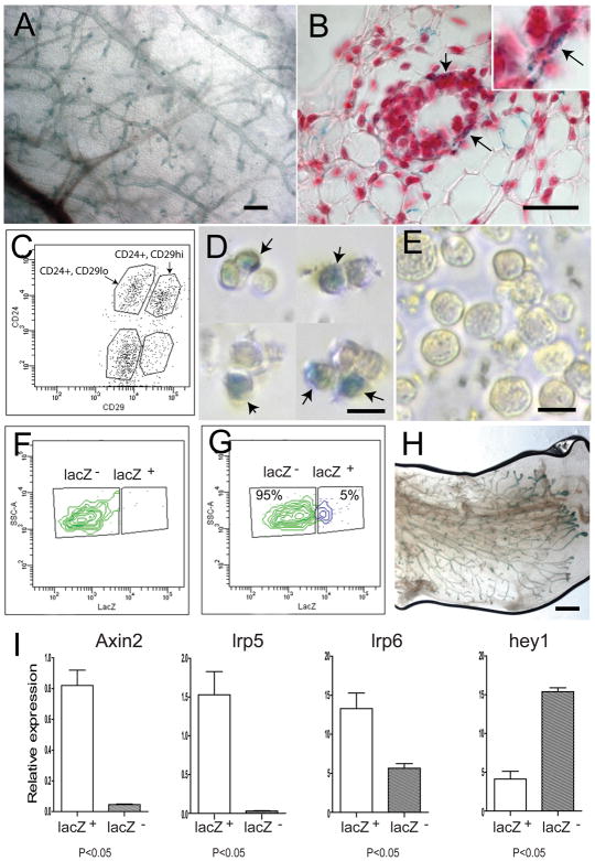
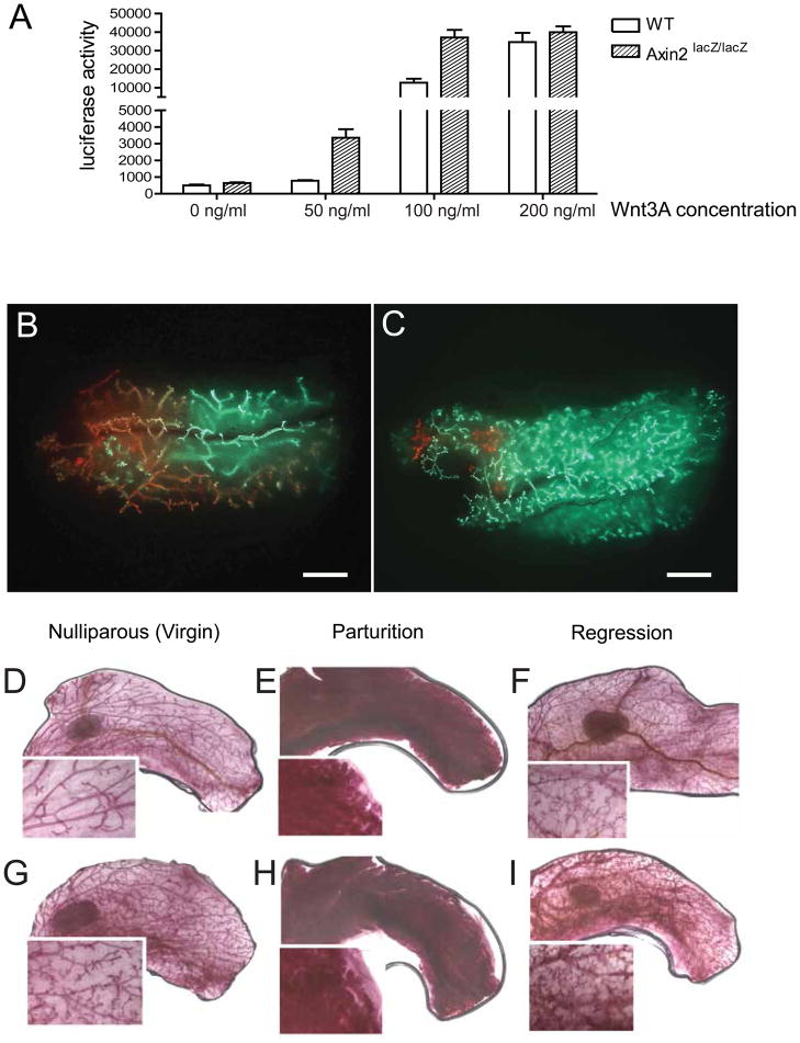
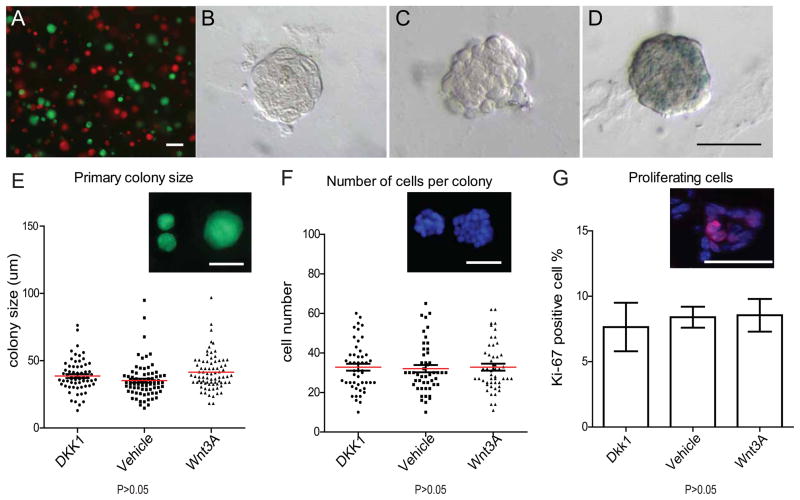
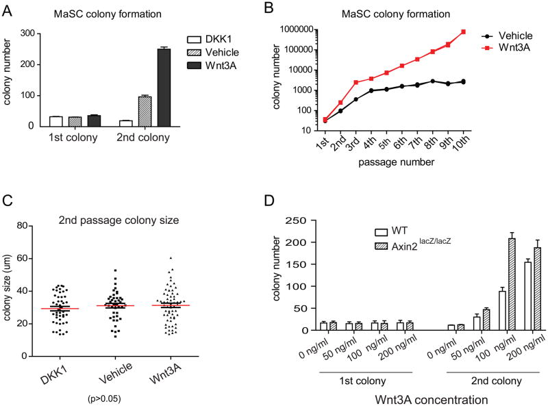
Adult stem cells have the ability to self-renew and to generate specialized cells. Self-renewal is dependent on extrinsic niche factors but few of those signals have been identified. In addition, stem cells tend to differentiate in the absence of the proper signals and are therefore difficult to maintain in cell culture. The mammary gland provides an excellent system to study self-renewal signals, because the organ develops postnatally, arises from stem cells, and is readily generated from transplanted cells. We show here that adult mammary glands contain a Wnt-responsive cell population that is enriched for stem cells. In addition, stem cells mutant for the negative-feedback regulator Axin2 and therefore sensitized to Wnt signals have a competitive advantage in mammary gland reconstitution assays. In cell culture experiments, exposure to purified Wnt protein clonally expands mammary stem cells for many generations and maintains their ability to generate functional glands in transplantation assays. We conclude that Wnt proteins serve as rate-limiting self-renewal signals acting directly on mammary stem cells.


Figures

Figure 1
Figure 1. The Wnt-responsive cell population is enriched for mammary stem cells
A, The expression of Axin2-lacZ was examined by whole-mount X-gal staining in Axin2+/lacZ virgin mammary gland (scale bars, 500um). Wnt-responsive cells are located in all branches. B, Paraffin sections revealed that Wnt-responsive cells (blue cells) reside in the basal layer of the mature ducts (arrows) (scale bar, 50um). Slides were counter-stained with nuclear fast red. C, Primary mammary epithelial cells (MECs) were isolated and analyzed by the expression of the cell surface markers lin, CD24 and CD29. D and E, lin, CD24+, CD29hi (stem cell enriched) and lin, CD24+, CD29low populations were FACS-isolated respectively, followed by X-gal staining. D, 5% of the lin, CD24+, CD29hi group is Wnt responsive (arrows; scale bar, 10um); E, no Wnt-responsive cells were seen in the lin, CD24+, CD29low group (scale bar, 10um). F, No lacZ+ cells were detected in the wild type cell control, using a LacZ fluorogenic substrate. G, 5% of lin, CD24+, CD29hicells are lacZ-positive in Axin2+/lacZ cells, using the lacZ fluorogenic substrate. H, Mammary outgrowths following transplantation were examined by X-gal staining to verify that the epithelium was donor cell derived (scale bar, 1mm). Using lacZ-expressing epithelial cells grown in wild type stroma, it is apparent that most if not all branches, contain Axin2-lacZ cells (compare 1H to 1A). I, q-RT-PCR relatively to HPRT control in the lacZ+, lin, CD24+, CD29hi and lacZ , lin, CD24+, CD29hi cells sorted from 8–12 week old virgin mouse mammary glands. A statistically significant difference of p<0.05 (t test) was observed between the two groups for each of the four genes.
Figure 2
Figure 2. Axin2lacZ/lacZ mutant cells have an advantage over wild type cells in competitive reconstitution assays
A, WT and Axin2lacZ/lacZ mammary epithelial cells transduced with a Wnt-reporter (7TCF-luciferase) construct were incubated with increasing concentrations of purified Wnt3A protein. The reporter activity was measured after 24 hours. B–C, Lin, CD24+, CD29hi cells were isolated independently from Actin-GFP mice and Actin-DsRed mice. GFP-positive and DsRed-positive cells were mixed in different ratios and injected into the cleared fat pads of recipients. As a control, B, transplantation of 1:1 ratio mixture of WT; Actin-DsRed and WT; Actin-GFP stem cells resulted in an even distribution of red and green outgrowths (scale bar, 2mm). C, More GFP-positive outgrowths were observed after transplantation of 2:1 mixture of Axin2+/+; DsRed and Axin2lacZ/lacZ ; Actin-GFP cells (scale bar, 2mm). D–F, Whole-mount carmine staining of WT mammary gland in nulliparous stage (D), parturition (E) and regression (F). Whole-mount carmine staining of Axin2lacZ/lacZ mammary gland. While Axin2lacZ/lacZ mammary glands in nulliparous stage display a mildly hypermorphic branching phenotype (G), they have normal differentiation in parturition (H) and regression in post-weaning (I).
Figure 3
Figure 3. Primary colony formation is not changed by Wnt signaling
A, FACS-isolated Lin, CD24+,CD29hi single cells were independently prepared from Actin-GFP mice or Actin-DsRed mice, mixed in a 1:1 ratio and seeded in Matrigel (2x10⁴ cells total in 50 ul pellet). Individual colonies that emerged were exclusively monochromatic, and the number of green and red colonies matched the ratio of the seeded cells (scale bars, 100um). B–D, Lin, CD24+, CD29hi cells isolated from Axin2lacZ/+ reporter mice were cultured in Matrigel, plus either Dkk1(B), Wnt vehicle control (C) or Wnt3A protein (D). X-gal staining was performed on colonies in 7 days of culture. No Wnt signaling was detectable in Dkk1 or vehicle controls, while the lacZ reporter was activated by Wnt3A protein (F) (scale bar 50um). E, Lin, CD24+,CD29hi cells isolated from Actin-GFP mice were cultured in Matrigel, plus either Dkk1, Wnt vehicle control or Wnt3A protein. Sizes of the colonies were measured after one week of culture in vitro. In all conditions, there are no differences in the average colony sizes, (which ranged from 20~60um, scale bar, 50um). F, DAPI staining of the colonies in E and cells numbers in each colony were quantified. G, Frozen sections of colonies in E were stained for the proliferation marker Ki-67. There was no difference in the number of Ki-67 positive cells among each condition (P>0.05).
Figure 4
Figure 4. In serial passage, Wnt3A protein leads to continued expansion of colony numbers
A. Lin, CD24+,CD29hi cells isolated from Actin-GFP mice were cultured in Matrigel, plus either Dkk1, Wnt vehicle control or Wnt3A protein. Numbers of primary colonies are similar (1 colony out of 15 plated cells in primary culture). However, the number of secondary colonies arising from re-plated single cells is strongly influenced by Wnt. In the presence of Dkk1, the secondary colony number dropped by 50%. In the presence of Wnt3A, the colony number was more than 7 fold higher. B, Colony numbers in vehicle and Wnt3A were followed over 10 passages. In vehicle, colony number reached a plateau in the 4th passage (black line). However, in the presence of Wnt3A, colony numbers expanded continually in each passage (red line). Starting from 35 primary colonies, 106 colonies were calculated to be present in the 10th passage. C, The sizes of the secondary colonies were measured after one week in culture post passage. Under each condition, there was no discernable difference in the average colony size. D. Primary and secondary colony numbers of wild type and Axin2lacZ/lacZ mutant cells grown over a range of Wnt3A protein concentration (X-axis). While primary colony formation efficiency remained unaffected, Axin2lacZ/lacZ mutant cells gave rise to higher numbers of secondary colonies compared to wild type cells (Y-axis), especially at limiting Wnt concentrations. At higher Wnt concentrations, the number of colonies plateaued irrespective of the genotype of the cells.
Figure 5
Figure 5. Cultured stem cell colonies retain full developmental potential
A, 100 GFP labeled Lin-,CD24+, CD29hi colonies cultured in the presence of Wnt3A for 2–6 passages were transplanted into cleared fat pads. Virgin recipients fat pads were harvested at 8 weeks post transplantation. Robust GFP outgrowths were detected. (scale bar, 2mm). B, C, Comparing H/E sections of WT (B) and reconstituted mammary glands (C) revealed a normal layered structure in the outgrowths (scale bars, 50um). D, An outgrowth from a transplanted single tertiary colony (scale bar, 2mm). E, F, Immuno-fluorescence staining with anti-K8 (E) and K14 antibody (F) of the sections indicated that single colony derived outgrowths have normal luminal (E) and myoepithelial (F) layers (scale bar, 50um). G, GFP outgrowths harvested from nursing recipients (day 2; scale bars, 2mm). H, Immunofluorescence staining with anti-milk antibody of alveoli arising from nursing recipients (Green, GFP epithelium; red, Milk; blue, DAPI. scale bar, 100um). I, GFP primary outgrowths have the same CD24 and CD29 profile as their donors. J, Normal secondary outgrowths arising from transplantation of cells isolated from primary outgrowths (scale bar, 2mm).

Comment in

References

    1. Al-Hajj M, Wicha MS, Benito-Hernandez A, Morrison SJ, Clarke MF. Prospective identification of tumorigenic breast cancer cells. Proc Natl Acad Sci U S A. 2003;100:3983–3988. - PMC - PubMed
    1. Asselin-Labat ML, Sutherland KD, Barker H, Thomas R, Shackleton M, Forrest NC, Hartley L, Robb L, Grosveld FG, van der Wees J, et al. Gata-3 is an essential regulator of mammary-gland morphogenesis and luminal-cell differentiation. Nat Cell Biol. 2007;9:201–209. - PubMed
    1. Badders NM, Goel S, Clark RJ, Klos KS, Kim S, Bafico A, Lindvall C, Williams BO, Alexander CM. The Wnt receptor, Lrp5, is expressed by mouse mammary stem cells and is required to maintain the basal lineage. PLoS One. 2009;4:e6594. - PMC - PubMed
    1. Baker R, Kent CV, Silbermann RA, Hassell JA, Young LJ, Howe LR. Pea3 transcription factors and wnt1-induced mouse mammary neoplasia. PLoS One. 2010;5:e8854. - PMC - PubMed
    1. Barker N, van Es JH, Kuipers J, Kujala P, van den Born M, Cozijnsen M, Haegebarth A, Korving J, Begthel H, Peters PJ, et al. Identification of stem cells in small intestine and colon by marker gene Lgr5. Nature. 2007;449:1003–1007. - PubMed

Publication types

MeSH terms