. 2010 Jun 4;6(6):578-90.
doi: 10.1016/j.stem.2010.04.002.

CNS-resident glial progenitor/stem cells produce Schwann cells as well as oligodendrocytes during repair of CNS demyelination

Affiliations

CNS-resident glial progenitor/stem cells produce Schwann cells as well as oligodendrocytes during repair of CNS demyelination

Malgorzata Zawadzka et al. Cell Stem Cell. .

Abstract

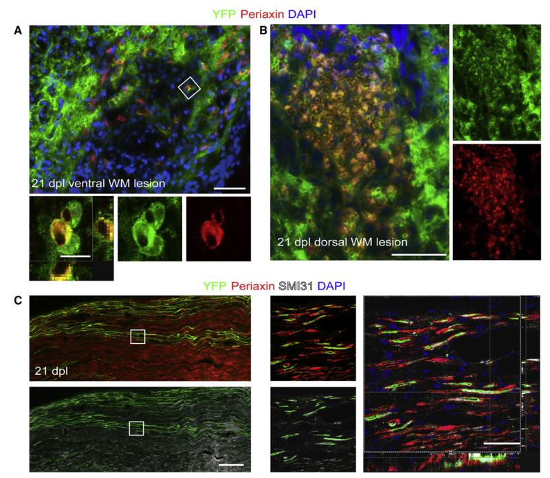
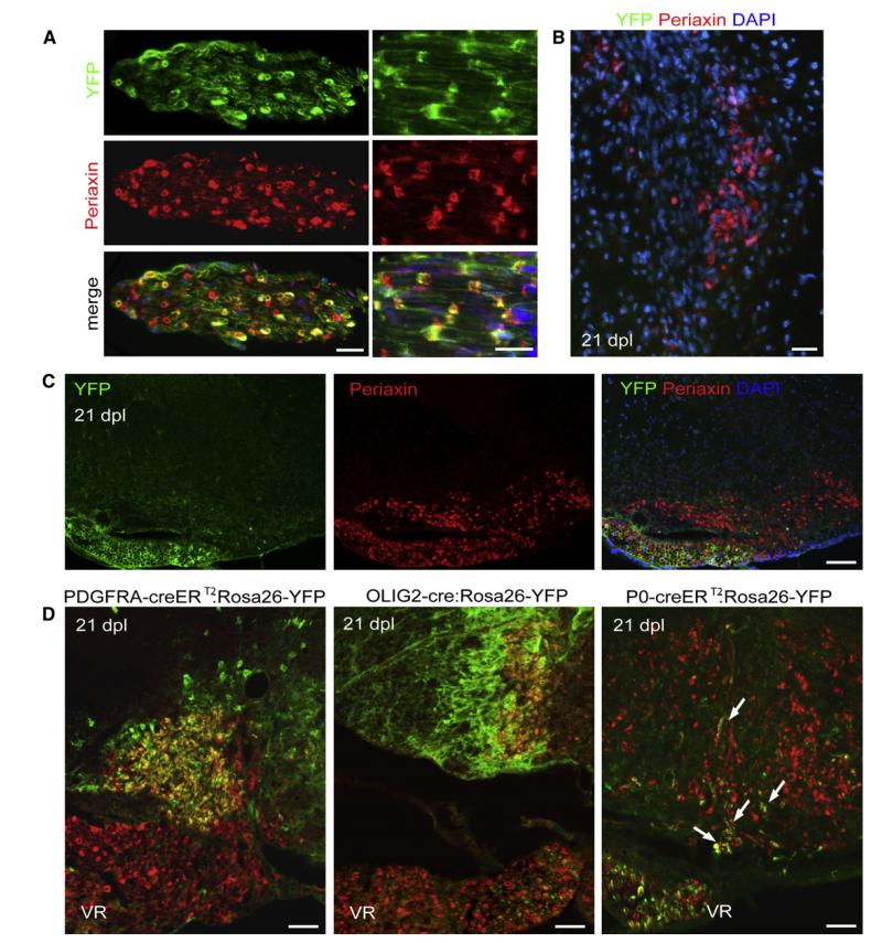
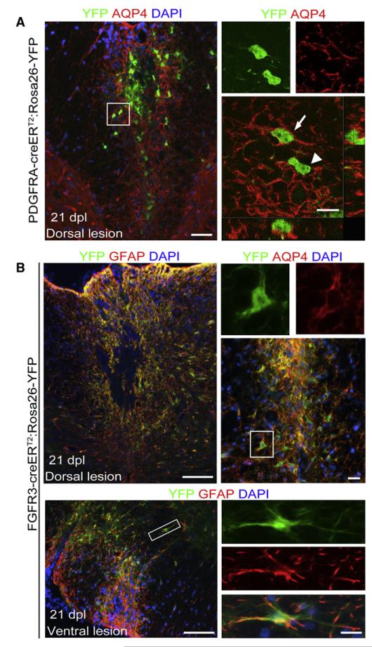
After central nervous system (CNS) demyelination-such as occurs during multiple sclerosis-there is often spontaneous regeneration of myelin sheaths, mainly by oligodendrocytes but also by Schwann cells. The origins of the remyelinating cells have not previously been established. We have used Cre-lox fate mapping in transgenic mice to show that PDGFRA/NG2-expressing glia, a distributed population of stem/progenitor cells in the adult CNS, produce the remyelinating oligodendrocytes and almost all of the Schwann cells in chemically induced demyelinated lesions. In contrast, the great majority of reactive astrocytes in the vicinity of the lesions are derived from preexisting FGFR3-expressing cells, likely to be astrocytes. These data resolve a long-running debate about the origins of the main players in CNS remyelination and reveal a surprising capacity of CNS precursors to generate Schwann cells, which normally develop from the embryonic neural crest and are restricted to the peripheral nervous system.


Figures

Figure 1
Figure 1. Identification of Genetically Labeled PDGFRA+ Cells in the Intact and Demyelinated Adult Spinal Cord
(A) Six days after induction of demyelination by injection of lysolecithin into the left ventrolateral white matter (dotted line), YFP+ cells can be seen throughout the white and gray matter and particularly concentrated within the lesion area (dpl, days postlesion; scale bar represents 500 μm). (B) Many of the YFP+ cells express OLIG2 in their nuclei (scale bars represent 100 μm in low-power image and 10 μm in higher-magnification confocal image). (C) Density of YFP+/OLIG2 and YFP+/OLIG2+ cells in normal nondemyelinated (intact) and demyelinated (lesion) tissue at 6, 14, and 21 DPL (n = 7, mean ± SD). (D) Nearly all YFP+ cells coexpress the OLP markers NG2 and PDGFRA (scale bars represent 20 μm).
Figure 2
Figure 2. Expression of Mature Oligodendrocyte Markers in YFP+ Cells with Morphological Features of Myelinating Oligodendrocytes
(A and B) Confocal images of spinal cord cross sections at 21 dpl immunostained with (A) anti-CC1 and (B) anti-Transferrin antibodies (scale bar represent 45 μm). High-power confocal projections of z-stacks show double-labeled cells from boxed regions (scale bars represent 10 μm). In the normal appearing white matter (NAWM), YFP+ cells do not express mature oligodendrocyte markers (scale bar, 20 μm). (C) Density of YFP+ cells expressing CC1 alone or CC1 and Transferrin at 14 and 21 dpl. (D) Longitudinal section of a remyelinated area at 21 dpl showing YPF+ processes closely associated with PLP+ myelin sheaths (arrows, scale bar represents 10 μm). (E) Alexa dye labeling of a YFP+ cell at 21 dpl by electroporation reveals a cell with a distinctive multiprocessed morphology characteristic of a myelinating oligodendrocyte (scale bar represents 20 μm, see also Movie S1).
Figure 3
Figure 3. Pdgfa-creERT2 Cells Give Rise to Myelinating Schwann Cells
(A) At 21 dpl, AQP-4 immunonegative (astrocyte-free) areas of the lesions can be identified containing YFP+/OLIG2-negative cells with morphologies resembling myelinating Schwann cells (left image) (the inset shows a group of cells in an adjacent section [corresponding to the boxed area], immunolabeled for OLIG2). Scale bars represent 100 μm (left image) and 20 μm (inset). Many of these YFP+ cells express the Schwann cell transcription factor SCIP within their nuclei (middle and right images, scale bar represents 10 μm). The histograms show the density of YFP+/SCIP and YFP+/SCIP+ cells at 14 and 21 dpl. (B) YFP+ cells with Schwann cell-like morphology are associated with Periaxin+ myelin sheaths (scale bar represents 50 μm). The boxed area in the left panel is shown at higher magnification in the middle panel and as a confocal Z-projection in the top right panel (scale bar represents 10 μm). The histograms show the density of YFP+/Periaxin and YFP+/Periaxin+ cells at 14 and 21 dpl. (C) Periaxin+ cells derived from PDGFRA+ precursors at 21 days after lesion show 1:1 interactions with two parallel SMI31+ axons in longitudinal section (indicated by arrowheads on merged image; scale bars represent 20 μm). (D) During remyelination of EB-induced lesions, a higher percentage of YFP+ cells coexpressed Periaxin+ than in lysolecithin-induced lesions at 21 dpl, while the proportion of remyelinating oligodendrocytes generated from PDGFRA+ precursors is approximately the same for both lesion types (p > 0.5, data expressed as percentage of YFP+ cells ± SEM).
Figure 4
Figure 4. PDGFRA Expression in Intact and Injured Sciatic Nerve of Pdgfra-creERT2 Mice
(A) YFP+ cells are detected at 6 days after nerve crush but not in intact noncrushed nerve; OLIG2 is not detected in either intact or crushed nerves. Longitudinal sections of sciatic nerves were double immuno-labeled for YFP and OLIG2 (scale bars represent 100 μm and 20 μm in low- and high-magnification images, respectively). RT-PCR analysis confirmed the lack of Olig2 transcripts in the intact or crushed sciatic nerve (lane 1, control intact nerve; lane 2, crushed nerve, 6 hr; lane 3, intact nerve, 24 hr; lane 4, crushed, 24 hr; lane 5, intact, 6 dpl; lane 6, crushed, 6 dpl; lane 7, dorsal root ganglia; compared with the level of expression in cultured OLPs; lanes 8 and 9, RNA from two separate OLP cultures), while Pdgfra can be detected in both intact and injured nerve (a housekeeping gene, Cyclophilin, was used as a normalization control). (B) Immunohistochemical characterization of YFP-expressing cells in 6 dpl sciatic nerve. YFP+ cells do not coexpress early Schwann cell markers such as SCIP, S100, and p75. Micrographs of longitudinal sections stained with anti-YFP and Schwann cell markers (main image scale bars represent 100 μm) and higher-magnification confocal projections of two different optical sections from the same field show nonoverlap between YFP and Schwann cell markers on any z-levels (inset scale bar represents 30 μm). (C and D) At 21 dpl some YFP+ cells could still be detected which strongly expressed fibronectin (D) but did not express Periaxin (C).
Figure 5
Figure 5. Remyelinating Schwann cells within CNS Lesion Derived from Olig2+ Cells
(A) Immunostaining for YFP and Periaxin performed on spinal cord cross sections from Olig2-cre: Rosa26-YFP mice at 21 days after lysolecithin-induced demyelination in ventral spinal cord white matter reveals cells expressing both markers (scale bar represents 50 μm) (confocal projection of cells from the boxed area, lower panel, scale bar represents 10 μm). (B) Overlay confocal image showing a high level of YFP/Periaxin colocalization (single channels for YFP and Periaxin; scale bar represents 50 μm) confirming that nearly all of Periaxin+ cells remyelinating lesions in the dorsal funiculus are generated from Olig2-cre-expressing cells. (C) YFP was expressed within sciatic nerve axons in Olig2-cre:Rosa26-YFP mice. There were no YFP+ cell bodies detected in injured sciatic nerves 28 days after nerve crush. Micrographs and confocal projections of longitudinal sections through the nerve show colocalization of YFP and SMI31 (axonal marker) but no overlap of YFP with the Schwann cell myelin protein Periaxin (scale bars represent 20 μm).
Figure 6
Figure 6. Some Periaxin+ Cells within Remyelinating CNS Lesions Are Derived from P0-Expressing Cells of the PNS, whereas the Majority Is Derived from PDGFRA- and OLIG2-Expressing CNS-Derived Cells
(A) Longitudinal and transverse sections of ventral root from P0-creERT2:Rosa26-YFP mice stained with anti-YFP and anti-Periaxin antibody show high efficiency of recombination 83% ± 2% (scale bars represent 30 μm). (B) No Periaxin+ cells generated from P0-creERT2-expressing cells are detected in the dorsal lysolecithin-induced lesions at 21 dpl (scale bars represent 30 μm). (C) Labeling of ventral roots in P0-creERT2:Rosa26-YFP mice provides an internal control for recombination efficiency. Although there is abundant Periaxin immunoreactivity in both the ventral roots and remyelinating lesions in the ventral funiculus at 21 dpl, YFP immunoreactivity is largely confined to the ventral root (scale bar represents 100 μm). (D) At 21 dpl in Pdgfra-creERT2:Rosa26-YFP or Olig2-Cre:Rosa26-YFP mice, there is extensive overlap of Periaxin and YFP immunoreactivity within remyelinating lesions but no overlap in the ventral roots (VR). The converse situation is found in P0-creERT2:Rosa26-YFP mice; there is very little overlap between Periaxin and YFP within demyelinated lesions (arrows) whereas nearly all Periaxin+ cells in the ventral roots are YFP+ (scale bars represent 50 μm).
Figure 7
Figure 7. PDGFRA+ Precursors Give Rise to Limited Number of Astrocytes in the Lesion, usually within the Outer Rim of the Lesion
(A) Low-power micrograph of a dorsal lesion immunolabelled for YFP and AQP4 (scale bar represents 50 μm). A cell with a YFP+ cytoplasm that also expresses AQP4 on its surface (arrow; scale bar represents 10 μm) can easily be distinguished from an AQP4-negative cell (arrowhead). (B) The majority of AQP4+ astrocytes within the lesion are derived from FGFR3+ cells as shown by double immunolabelling for YFP and either GFAP or AQP4 in dorsal or ventral lesions (scale bars represent 100 μm in main image, 20 μm in higher-magnification images of areas indicated by boxes).

References

    1. Arnett HA, Fancy SPJ, Alberta JA, Zhao C, Plant SR, Kaing S, Raine CS, Rowitch DH, Franklin RJ, Stiles CD. bHLH transcription factor Olig1 is required to repair demyelinated lesions in the CNS. Science. 2004;306:2111–2115. - PubMed
    1. Baron-Van Evercooren A, Avellana-Adalid V, Ben Younes-Chennoufi A, Gansmuller A, Nait-Oumesmar B, Vignais L. Cell-cell interactions during the migration of myelin-forming cells transplanted in the demyelinated spinal cord. Glia. 1996;16:147–164. - PubMed
    1. Belachew S, Chittajallu R, Aguirre AA, Yuan X, Kirby M, Anderson S, Gallo V. Postnatal NG2 proteoglycan-expressing progenitor cells are intrinsically multipotent and generate functional neurons. J. Cell Biol. 2003;161:169–186. - PMC - PubMed
    1. Blakemore WF. Remyelination by Schwann cells of axons demyelinated by intraspinal injection of 6-aminonicotinamide in the rat. J. Neurocytol. 1975;4:745–757. - PubMed
    1. Blakemore WF, Franklin RJM. Remyelination in experimental models of toxin-induced demyelination. Curr. Top. Microbiol. Immunol. 2008;318:193–212. - PubMed

Publication types

MeSH terms

Substances