Comparative Study
. 2010 Jun 4;6(6):591-602.
doi: 10.1016/j.stem.2010.03.016.

Wilms tumor chromatin profiles highlight stem cell properties and a renal developmental network

Affiliations
Comparative Study

Wilms tumor chromatin profiles highlight stem cell properties and a renal developmental network

Aviva Presser Aiden et al. Cell Stem Cell. .

Abstract

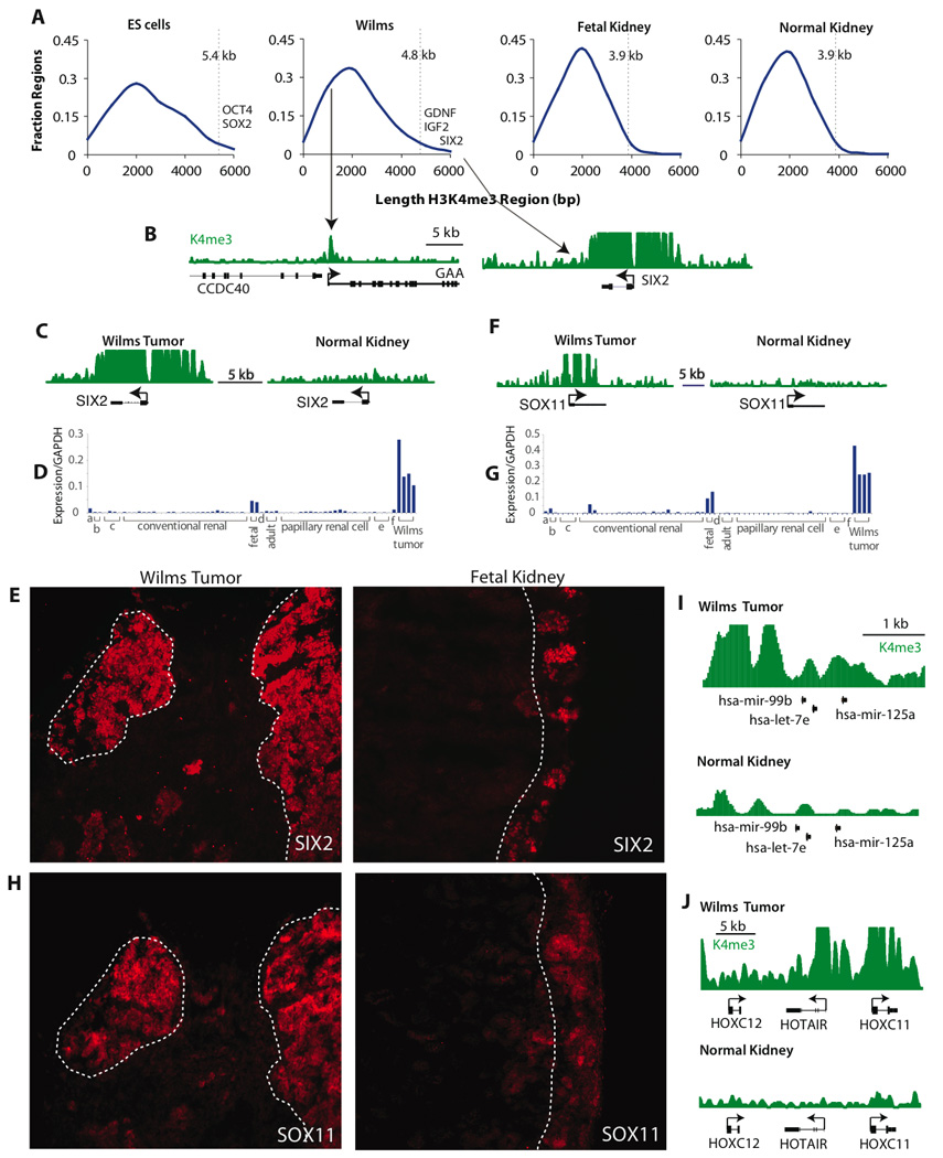
Wilms tumor is the most common pediatric kidney cancer. To identify transcriptional and epigenetic mechanisms that drive this disease, we compared genome-wide chromatin profiles of Wilms tumors, embryonic stem cells (ESCs), and normal kidney. Wilms tumors prominently exhibit large active chromatin domains previously observed in ESCs. In the cancer, these domains frequently correspond to genes that are critical for kidney development and expressed in the renal stem cell compartment. Wilms cells also express "embryonic" chromatin regulators and maintain stem cell-like p16 silencing. Finally, Wilms and ESCs both exhibit "bivalent" chromatin modifications at silent promoters that may be poised for activation. In Wilms tumor, bivalent promoters correlate to genes expressed in specific kidney compartments and point to a kidney-specific differentiation program arrested at an early-progenitor stage. We suggest that Wilms cells share a transcriptional and epigenetic landscape with a normal renal stem cell, which is inherently susceptible to transformation and may represent a cell of origin for this disease.


Figures

Figure 1
Figure 1. Gene classification based on chromatin provides insights into the developmental state of Wilms tumor
(a) Wilms tumors, normal kidney and ES cells were clustered by K4me3 promoter states. The cluster tree depicts relationships among these different tissues. (b, c) Four gene sets were defined based on promoter chromatin states. For each set, chromatin profiles are shown for a typical gene. These profiles depict ChIP-Seq signals for the indicated modification. The x-axis corresponds to genome position. The y-axis corresponds to the number of sequenced ChIP fragments that overlap a given position (range = 0–12). Set 1 contains genes with broad K4me3 domains in Wilms tumor; it is enriched for known regulators of kidney and mesoderm development (top two categories shown). Set 2 contains genes with promoter K4me3 peaks in Wilms tumor and ES cells, but not in normal kidney; it is enriched for regulators of chromatin, transcription and epigenetic processes. Set 3 contains genes with overlapping K4me3 and K27me3, a subset of which is differentially-expressed in kidney (see text and Fig 3). Set 4 contains genes with promoter K27me3 peaks; it is enriched for genes involved in distinct developmental processes. See also Fig S1, Tables S1–S5.
Figure 2
Figure 2. Domains of modified histones identify developmental regulators in Wilms tumor
(a) Histograms depict the size distribution of K4me3 enriched regions in ES cells, Wilms tumor, fetal kidney and normal kidney. In both ES cells and Wilms tumor, the most expansive regions correspond to developmental genes with critical roles in the respective cell types. 95th percentile values (5.4 kb for ES cells, 4.8 for Wilms tumor, and 3.9 kb for both fetal and normal kidney) are indicated on each plot. (b) Genomic views show K4me3 signal in Wilms tumor for a typical promoter (GAA) and for the kidney stem cell regulator SIX2. (c–d) K4me3 signal at the SIX2 and SOX11 loci in Wilms tumor compared to normal kidney. (e–f) Expression of SIX2 and SOX11 mRNA shown for a panel of renal tumors and tissues [a: clear cell sarcoma of the kidney; b: collecting duct carcinoma; c: chromophobe renal cell cancer; conventional renal cell carcinoma; fetal kidney; d: renal lipoma; adult kidney; papillary renal cell carcinoma; e: renal oncocytoma; f: rhabdoid tumor of kidney; Wilms tumor]. (g–h) Immunofluorescence images depict SIX2 and SOX11 protein expression in a WTX mutant Wilms tumor and fetal kidney. In Wilms tumor, SIX2 and SOX11 have similar expression in the blastemal compartment (dotted outlines). In fetal kidney both markers are adjacent to the surface of the organ where mesenchymal and epithelial precursors are located (dotted lines). Original magnification 200×. (i,j) Genomic views show non-coding RNA genes with broad K4me3 domains in Wilms tumor. See also Tables S6,S7.
Figure 3
Figure 3. Gene sets defined by chromatin state in Wilms tumor show differential expression patterns in normal and malignant kidney tissues
Row-normalized heat maps depict relative expression of genes in Set 1 and Set 3. Red indicates high levels of expression; blue indicates low or no expression. (a) Genes in Set 1 (broad K4me3) are preferentially expressed in Wilms tumor and fetal kidney (Yusenko et al., 2009). (b) Genes in Set 3 show relatively greater expression in adult kidney, and heterogeneous expression in conventional renal cell carcinoma (cRCC). See also Fig S2.
Figure 4
Figure 4. Chromatin regulators in Wilms tumor and ES cells
(a) Genomic views depict K4me3 and K27me3 signals for genes encoding key chromatin regulators that are common to Wilms tumor and ES cells, but inactive in normal kidney and adult renal cancers. The highlighted genes are a subset of ‘embryonic’ chromatin regulators active in Wilms tumor. See also Fig S3, Table S8, S9.
Figure 5
Figure 5. Protein expression patterns of bivalent genes in Wilms tumor and kidney tissue
Genomic views depict K4me3 and K27me3 signals for (a) KCNJ3 and (b) NR4A2 which are bivalent in the tumor cells (top panels). Variable transition to a more active chromatin state is evident in normal kidney which is heterogeneous. Immunofluorescence images show expression of the corresponding proteins in Wilms tumor and normal kidney (bottom panels). The images reveal weak expression in tumor, consistent with the chromatin patterns, but strong staining of specific epithelial compartments of normal kidney (red signals). Nuclei are stained with DAPI (blue signal). Original magnification 400×. See also Fig S4.
Figure 6
Figure 6. Epigenetic regulation in Wilms tumor
(a) Genomic view depicts K4me3 and K27me3 signals for the p16 tumor suppressor locus (CDKN2A). This locus remains intact in the tumor, but is silenced by expansive K27me3 in a pattern reminiscent of normal stem cells. (b) Methylation Specific PCR (MSP) shows that the p16 locus remains unmethylated at the DNA level, a pattern typical of normal stem cells but distinct from many adult cancers. Bisulfite converted DNA was amplified using primers specific for unmethylated (p16 U) or methylated (p16 M) versions of the locus. Gels show PCR products for unmethylated (U) and methylated (M) genomic DNA controls and three Wilms tumor samples. (c) Genomic views depict K4me3 and K27me3 signals for imprinted gene loci. IGF2, which has been causally implicated in Wilms tumor, is associated with a uniquely broad K4me3 domain in Wilms tumor. PEG3 also shows substantially more K4me3 in the tumor.

References

    1. Azuara V, Perry P, Sauer S, Spivakov M, Jorgensen HF, John RM, Gouti M, Casanova M, Warnes G, Merkenschlager M, et al. Chromatin signatures of pluripotent cell lines. Nat Cell Biol. 2006;8:532–538. - PubMed
    1. Bardeesy N, Falkoff D, Petruzzi MJ, Nowak N, Zabel B, Adam M, Aguiar MC, Grundy P, Shows T, Pelletier J. Anaplastic Wilms' tumour, a subtype displaying poor prognosis, harbours p53 gene mutations. Nat Genet. 1994;7:91–97. - PubMed
    1. Barski A, Cuddapah S, Cui K, Roh TY, Schones DE, Wang Z, Wei G, Chepelev I, Zhao K. High-resolution profiling of histone methylations in the human genome. Cell. 2007;129:823–837. - PubMed
    1. Beckwith JB, Kiviat NB, Bonadio JF. Nephrogenic rests, nephroblastomatosis, and the pathogenesis of Wilms' tumor. Pediatr Pathol. 1990;10:1–36. - PubMed
    1. Ben-Porath I, Thomson MW, Carey VJ, Ge R, Bell GW, Regev A, Weinberg RA. An embryonic stem cell-like gene expression signature in poorly differentiated aggressive human tumors. Nat Genet. 2008;40:499–507. - PMC - PubMed

Publication types

MeSH terms

Associated data