

Review
doi: 10.1016/j.ceb.2010.05.005.
Epub 2010 Jun 1.
Quality and quantity control at the endoplasmic reticulum
Affiliations
- PMID: 20570125
- PMCID: PMC2929805
- DOI: 10.1016/j.ceb.2010.05.005
Item in Clipboard
Review
Quality and quantity control at the endoplasmic reticulum
Curr Opin Cell Biol.
2010 Aug.
Abstract
The endoplasmic reticulum (ER) is the site of maturation for secretory and membrane proteins that together make up about one third of the cellular proteome. Cells carefully control the synthetic output of this organelle to regulate both quality and quantity of proteins that emerge. Here, we synthesize current concepts underlying the pathways that mediate protein degradation from the ER and their deployment under physiologic and pathologic conditions.
Published by Elsevier Ltd.
Figures
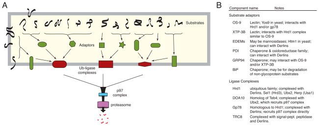
(A) General pyramidal scheme with many substrates, several adaptors, a handful of membrane complexes, and a commonly shared mechanism for substrate extraction and degradation in the cytosol. Substrates vary with regard to topology, post-translational modifications, and nature of the folding defect. These parameters influence the specific pathway(s) available to the substrate. Although not depicted, some substrates might engage a ubiquitin ligase complex directly. There may also be considerable overlap among pathways: substrates could access multiple adaptors, and adaptors might be capable of binding multiple ligase complexes. (B) Several examples of putative adaptors (many of which are chaperones) and ubiquitin ligase complex components are listed.
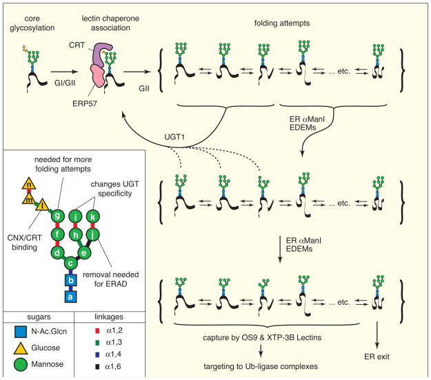
Newly synthesized proteins are core-glycosylated (upper left) with a highly asymmetric 14-hexose glycan (see inset for details). The glucoses are trimmed by glucosidase I and glucosidase II (GI/GII), generating a mono-glucosylated glycan that binds Calreticulin (CRT) or Calnexin, along with an associated oxidoreductase such as ERP57. Upon release, the terminal glucose can be trimmed by GII, preventing re-binding by CRT. During this time, the substrate accesses various possible folding conformations. Depending on the conformation, the substrate can be acted upon by either UGT1 (which re-glucosylates the glycan) or ER mannosidases such as αER-ManI and possibly EDEM family members. Mannose-trimmed glycans can still potentially be re-glucosylated by UGT1 (albeit with lower efficiency) or further de-mannosylated, depending again on the folding status. Removal of the ‘g’ mannose (see inset) irreversibly precludes re-glucosylation, precluding any further folding attempts. The substrate then only has the option of degradation or ER exit. Depending on its folding state, it is thought mannosidases like EDEM family members remove the ‘k’ mannose, exposing the α1,6 linked ‘j’ mannose needed for binding the lectin ERAD adaptors OS9 or XTP-3B. Other lectins such as ERGIC53 facilitate ER export. Note that many substrates have multiple glycans and multiple folding domains, markedly increasing the complexity of these reactions. Note that the precise glycan structures generated by each enzyme and recognized by the different lectins remains to be fully elucidated.
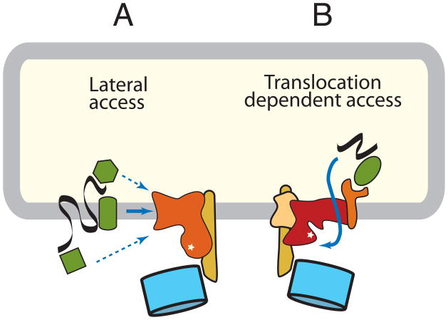
Membrane ubiquitin ligase complexes mediate substrate access to the catalytic site by two distinct mechanisms. (A) Membrane protein substrates might access the catalytic site by lateral delivery. Recognition and targeting might be mediated by an adaptor in the membrane, cytosol, or lumen. Alternatively, the ubiquitin ligase complex itself could recognize some substrates. (B) Lumenal substrates and some membrane proteins access the catalytic site by a translocation-dependent mechanism. The mechanism or components mediating the key translocation step to provide initial substrate access is unknown, but might involve the ubiquitin ligase itself or an associated membrane protein. In yeast, a complex centered around the Doa10 ubiquitin ligase is probably an example of the first pathway, while a complex containing the Hrd1 ubiquitin ligase is an example of the second pathway. In mammals, many additional similar complexes built around other ubiquitin ligases exist, although their compositions remain to be clearly defined.
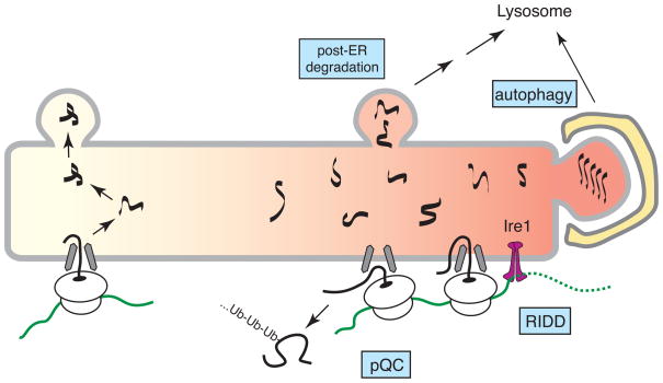
During particularly severe ER stress, several pathways of quality control that may not operate during normal conditions become important for limiting protein misfolding in the ER. Pre-emptive quality control (pQC) involves reduced translocation of certain protein that are instead routed into the cytosol for degradation. Regulated Ire1-dependent degradation (RIDD) mediates degradation of select ER-bound mRNAs. Some misfolded proteins may be degraded by post-ER pathways involving vesicular trafficking to the lysosome. Autophagy can sequester whole sections of the ER containing misfolded or aggregated proteins.
References
Publication types
MeSH terms
Grants and funding
LinkOut - more resources
Full Text Sources
Other Literature Sources
