

Automated MRI measures predict progression to Alzheimer's disease
- PMID: 20570399
- PMCID: PMC2902697
- DOI: 10.1016/j.neurobiolaging.2010.04.023
Automated MRI measures predict progression to Alzheimer's disease
Abstract
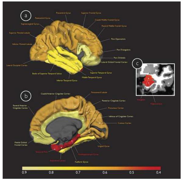
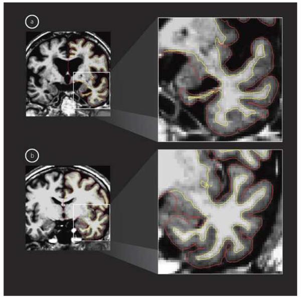
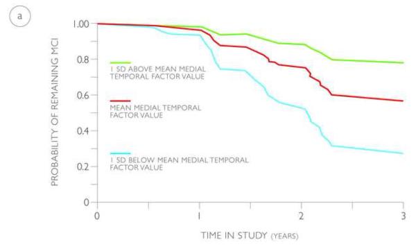
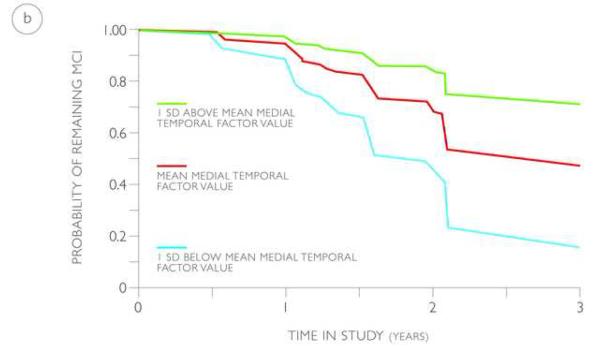
The prediction of individuals with mild cognitive impairment (MCI) destined to develop Alzheimer's disease (AD) is of increasing clinical importance. In this study, using baseline T1-weighted MRI scans of 324 MCI individuals from two cohorts and automated software tools, we employed factor analyses and Cox proportional hazards models to identify a set of neuroanatomic measures that best predicted the time to progress from MCI to AD. For comparison, cerebrospinal fluid (CSF) assessments of cellular pathology and positron emission tomography (PET) measures of metabolic activity were additionally examined. By 3 years follow-up, 60 MCI individuals from the first cohort and 58 MCI individuals from the second cohort had progressed to a diagnosis of AD. Cox models on the first cohort demonstrated significant effects for the medial temporal factor [Hazards Ratio (HR) = 0.43{95% confidence interval (CI), 0.32-0.55}, p < 0.0001], the fronto-parietoccipital factor [HR = 0.59{95% CI, 0.48-0.80}, p < 0.001], and the lateral temporal factor [HR = 0.67 {95% CI, 0.52-0.87}, p < 0.01]. When applied to the second cohort, these Cox models showed significant effects for the medial temporal factor [HR = 0.44 {0.32-0.61}, p < 0.001] and lateral temporal factor [HR = 0.49 {0.38-0.62}, p < 0.001]. In a combined Cox model, consisting of individual CSF, PET, and MRI measures that best predicted disease progression, only the medial temporal factor [HR = 0.53 {95% CI, 0.34-0.81}, p < 0.001] demonstrated a significant effect. These findings illustrate that automated MRI measures of the medial temporal cortex accurately and reliably predict time to disease progression, outperform cellular and metabolic measures as predictors of clinical decline, and can potentially serve as a predictive marker for AD.
Elsevier Inc. All rights reserved.
Figures
References
-
- Apostolova LG, Dutton RA, Dinov ID, Hayashi KM, Toga AW, Cummings JL, Thompson PM. Conversion of mild cognitive impairment to Alzheimer disease predicted by hippocampal atrophy maps. Arch. Neurol. 2006;63:693–699. - PubMed
-
- Arnold SE, Hyman BT, Flory J, Damasio AR, Van Hoesen GW. The topographical and neuroanatomical distribution of neurofibrillary tangles and neuritic plaques in the cerebral cortex of patients with Alzheimer's disease. Cereb. Cortex. 1991;1:103–116. - PubMed
-
- Attwell D, Laughlin SB. An energy budget for signaling in the grey matter of the brain. J. Cereb. Blood. Flow. Metab. 2001;21:1133–1145. - PubMed
-
- Braak H, Braak E. Neuropathological stageing of Alzheimer-related changes. Acta Neuropathology (Berlin) 1991;82:239–259. - PubMed
Publication types
MeSH terms
Grants and funding
- U24 RR021382/RR/NCRR NIH HHS/United States
- P50 AG05681/AG/NIA NIH HHS/United States
- P41-RR14075/RR/NCRR NIH HHS/United States
- R01 RR 16594-01A1/RR/NCRR NIH HHS/United States
- R01 EB001550/EB/NIBIB NIH HHS/United States
- AG021910/AG/NIA NIH HHS/United States
- R01 AG021910/AG/NIA NIH HHS/United States
- R01 RR016594/RR/NCRR NIH HHS/United States
- P41 RR006009/RR/NCRR NIH HHS/United States
- R01 NR010827/NR/NINR NIH HHS/United States
- U01 AG024904/AG/NIA NIH HHS/United States
- U19 AG010483/AG/NIA NIH HHS/United States
- P01 AG003991/AG/NIA NIH HHS/United States
- P50 AG005681/AG/NIA NIH HHS/United States
- P01 AG03991/AG/NIA NIH HHS/United States
- P41 RR014075/RR/NCRR NIH HHS/United States
LinkOut - more resources
Full Text Sources
Other Literature Sources
Medical
