. 2010 Sep;31(5):485-92.
doi: 10.1016/j.neuro.2010.05.014. Epub 2010 Jun 8.

Higher susceptibility of the ventral versus the dorsal hippocampus and the posteroventral versus anterodorsal amygdala to soman-induced neuropathology

Affiliations

Higher susceptibility of the ventral versus the dorsal hippocampus and the posteroventral versus anterodorsal amygdala to soman-induced neuropathology

James P Apland et al. Neurotoxicology. 2010 Sep.

Abstract

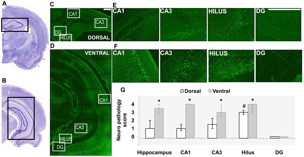
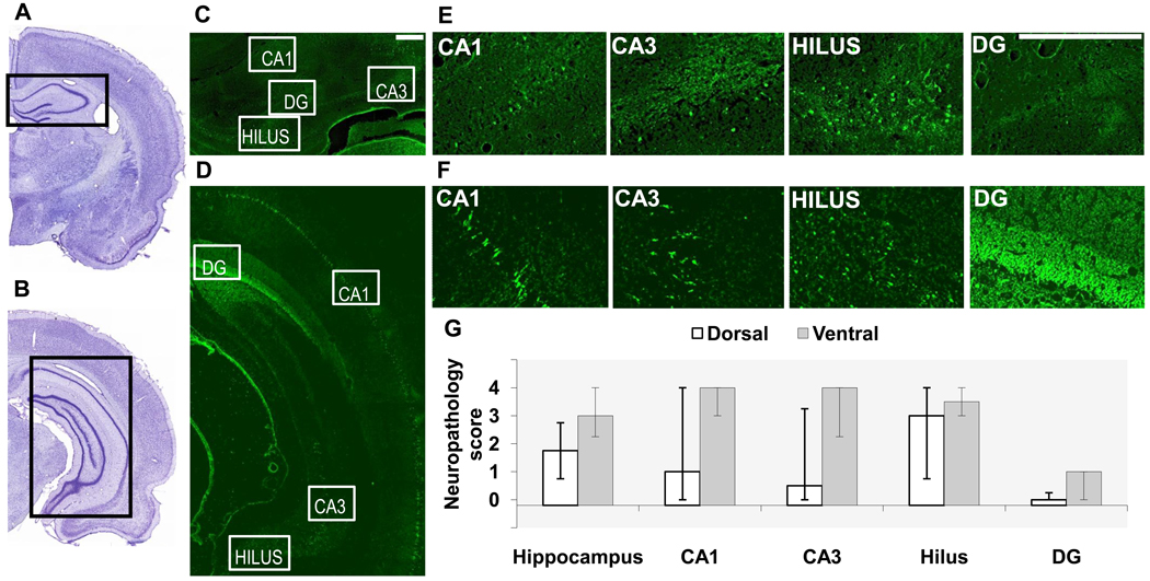
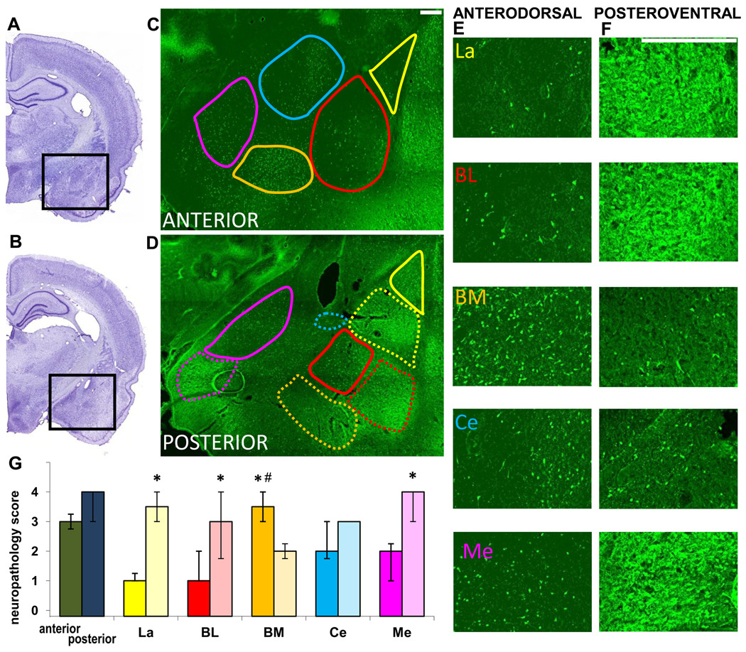
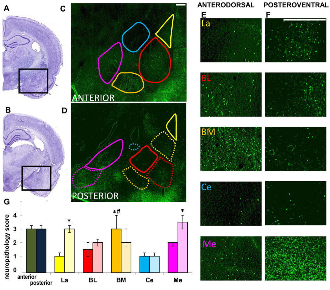
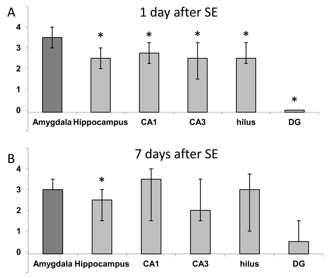
Nerve agents are acetylcholinesterase inhibitors, exposure to which causes brain damage, primarily by inducing intense seizure activity. Knowledge of the brain regions that are most vulnerable to nerve agent-induced brain damage can facilitate the development of drugs targeting the protection of these regions. Both the amygdala and the hippocampus have been shown to suffer significant damage after nerve agent exposure, but the amygdala appears to be the more severely affected structure. However, damage in the amygdala has generally been compared with damage in the dorsal hippocampus, whereas there is evidence that the ventral hippocampus is significantly more susceptible to seizures than the dorsal region and, therefore, it may also be more susceptible to nerve agent-induced neuropathology. Here, we report that after status epilepticus induced by soman administration to rats, neuronal degeneration as assessed by Fluoro-Jade C staining was more extensive in the ventral than the dorsal hippocampal subfields, 1 day after soman exposure. Seven days later, the difference between dorsal and ventral regions was not statistically significant. In the amygdala, soman-induced neurodegeneration was more severe in the posteroventral regions of the lateral, basolateral, and medial nuclei compared to the anterodorsal regions of these nuclei. In contrast, the basomedial nucleus was more severely affected in the anterodorsal region. The extent of neurodegeneration in the amygdala was not significantly different from that in the ventral hippocampus. However, when compared with the whole hippocampus, the amygdala displayed more severe neurodegeneration, on both day 1 and day 7 after soman exposure. Testing the protective efficacy of drugs against nerve agent-induced brain damage should include examination of the ventral hippocampus and the posteroventral regions of the amygdala, as these areas are most vulnerable to nerve agent-induced neurodegeneration.


Conflict of interest statement

Conflict of Interest statement: The authors declare that there are no conflicts of interest.

Figures

Fig. 1
Fig. 1. Neuronal degeneration is greater in the ventral hippocampus compared to the dorsal hippocampus, 1 day after soman-induced SE
A, B. Photomicrographs of panoramic Nissl-stained coronal brain sections showing the dorsal (A) and ventral (B) hippocampus. C, D, E, F. Photomicrographs of Fluoro-Jade C-stained sections showing the dorsal (C, E) and ventral (D, F) hippocampal subfields (scale bar is 300µ for both magnifications). G. Bar graph showing medians and interquartile range (IQR) of the neuropathology scores for the whole hippocampus, CA1 and CA3 subfields, hilar region, and granule cell layer of the dentate gyrus (IQR bar is not present in the CA1 area of the ventral hippocampus because there was no variability among animals in the neuropathology score). *p < 0.05 for comparisons between dorsal and ventral regions. #p < 0.05 for comparisons between the dorsal hilus and the other dorsal hippocampal subfields.
Fig. 2
Fig. 2. Neuronal degeneration in the dorsal and ventral hippocampus, 7 days after soman-induced SE
A, B. Photomicrographs of panoramic Nissl-stained coronal brain sections showing the dorsal (A) and ventral (B) hippocampus. C, D, E, F. Photomicrographs of Fluoro-Jade C-stained sections showing the dorsal (C, E) and ventral (D, F) hippocampal subfields (scale bar is 300µ for both magnifications). G. Bar graph showing medians and interquartile range of the neuropathology scores for the whole hippocampus, CA1 and CA3 subfields, hilar region, and granule cell layer of the dentate gyrus (DG).
Fig. 3
Fig. 3. Neuronal degeneration in amygdalar nuclei, 1 day after soman-induced SE
A, B. Photomicrographs of panoramic Nissl-stained coronal brain sections showing the anterior (A) and posterior (B) amygdala. C, D. Photomicrographs of Fluoro-Jade C-stained sections of the amygdala, outlining the studied nuclei in the anterior and posterior amygdala (La-yellow; BL-red; BM-orange; Ce-blue; Me-purple). Contours are traced in solid lines for the anterior and dorsal (“anterodorsal”) subdivisions and in dashed lines for the posterior and ventral (“posteroventral”) subdivisions. E, F. Photomicrographs of Fluoro-Jade C-stained sections showing the different anterodorsal and posteroventral amygdala nuclei (scale bar is 300µ for both magnifications). G. Bar graph showing medians and interquartile range of the neuropathology scores for the anterior and posterior sections of the amygdala, and individual amygdala nuclei (solid colors for the anterodorsal and transparent colors for the posteroventral nuclei). *p < 0.05 for comparisons between the anterodorsal and posteroventral regions. #p < 0.05 for comparisons between the anterodorsal BM nucleus and the other anterodorsal nuclei.
Fig. 4
Fig. 4. Neuronal degeneration in amygdalar nuclei, 7 days after soman-induced SE
A, B. Photomicrographs of panoramic Nissl-stained coronal brain sections showing the anterior (A) and posterior (B) amygdala. C, D. Photomicrographs of Fluoro-Jade C-stained sections of the amygdala, outlining the studied nuclei in the anterior and posterior amygdala (La-yellow; BL-red; BM-orange; Ce-blue; Me-purple). Contours are traced in solid lines for the anterior and dorsal (“anterodorsal”) subdivisions and in dashed lines for the posterior and ventral (“posteroventral”) subdivisions. E, F. Photomicrographs of Fluoro-Jade C-stained sections showing the different anterodorsal and posteroventral amygdala nuclei (scale bar is 300µ for both magnifications). G. Bar graph showing medians and interquartile range of the neuropathology scores for the anterior and posterior sections of the amygdala, and individual amygdala nuclei (solid colors for the anterodorsal and transparent colors for the posteroventral). *p < 0.05 for comparisons between the anterodorsal and posteroventral regions. #p < 0.05 for comparisons between the anterodorsal BM nucleus and the other anterodorsal nuclei.
Fig. 5
Fig. 5. Neuronal degeneration in the amygdala is significantly greater than that in the hippocampus, after soman-induced SE
When the whole amygdala and the whole hippocampus were compared (average neurodegeneration score from sections along the anterodorsal to posteroventral axis), the amygdala displayed greater damage than the hippocampus, on both day 1 (A) and day 7 (B) after SE. Amygdala damage was also greater when compared with individual hippocampal subfields along the extent of the hippocampus, on day 1 but not on day 7 after SE. The bars show the medians and interquartile range. Asterisks indicating statistical significance (p < 0.05) have been placed on the bars that are statistically lower than the bar depicting amygdala neuropathology.

References

    1. Akaike K, Tanaka S, Hideshi Tojo H, Fukumoto S, Imamura S, Takigawa M. Kainic acid-induced dorsal and ventral hippocampal seizures in rats. Brain Res. 2001;900:65–71. - PubMed
    1. Apland JP, Aroniadou-Anderjaska V, Braga MFM. Soman induces ictogenesis in the amygdala and interictal activity in the hippocampus that are blocked by a GluR5 kainate receptor antagonist in vitro. Neuroscience. 2009;159:380–389. - PMC - PubMed
    1. Aroniadou-Anderjaska V, Figueiredo YH, Apland JP, Qashu F, Braga MFM. Primary brain targets of nerve agents: the role of the amygdala in comparison to the hippocampus. Neurotoxicology. 2009 In Press. - PMC - PubMed
    1. Baille V, Clarke PG, Brochier G, Dorandeu F, Verna JM, Four E, Lallement G, Carpentier P. Soman-induced convulsions: the neuropathology revisited. Toxicology. 2005;215:1–24. - PubMed
    1. Bajgar J, Sevelova L, Krejcova G, Fusek J, Vachek J, Kassa J, Herink J, de Jong LP, Benschop HP. Biochemical and behavioral effects of soman vapors in low concentrations. Inhal Toxicol. 2004;16:497–507. - PubMed

Publication types

MeSH terms

LinkOut - more resources