. 2010 Oct;38(10):896-907.
doi: 10.1016/j.exphem.2010.05.008. Epub 2010 Jun 1.

H1-receptor antagonists terfenadine and loratadine inhibit spontaneous growth of neoplastic mast cells

Affiliations

H1-receptor antagonists terfenadine and loratadine inhibit spontaneous growth of neoplastic mast cells

Emir Hadzijusufovic et al. Exp Hematol. 2010 Oct.

Abstract

Objective: In mast cell (MC) neoplasms, clinical problems requiring therapy include local aggressive and sometimes devastating growth of MCs and mediator-related symptoms. A key mediator of MCs responsible for clinical symptoms is histamine. Therefore, use of histamine receptor (HR) antagonists is an established approach to block histamine effects in these patients.

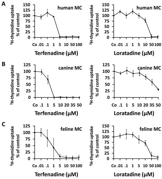
Materials and methods: We screened for additional beneficial effects of HR antagonists and asked whether any of these agents would also exert growth-inhibitory effects on primary neoplastic MCs, the human MC line HMC-1, and on two canine MC lines, C2 and NI-1.

Results: We found that the HR1 antagonists terfenadine and loratadine suppress spontaneous growth of HMC-1, C2, and NI-1 cells, as well as growth of primary neoplastic MCs in all donors tested (human patients, n = 5; canine patients, n = 8). The effects of both drugs were found to be dose-dependent (IC(50): terfenadine, 1-20 μM; loratadine, 10-50 μM). Both agents also produced apoptosis in neoplastic MCs and augmented apoptosis-inducing effects of two KIT-targeting drugs, PKC412 and dasatinib. The other HR1 antagonists (fexofenadine, diphenhydramine) and HR2 antagonists (famotidine, cimetidine, ranitidine) tested did not exert substantial growth-inhibitory effects on neoplastic MCs. None of the histamine receptor blockers were found to modulate cell-cycle progression in neoplastic MCs.

Conclusions: The HR1 antagonists terfenadine and loratadine, in addition to their antimediator activity, exert in vitro growth-inhibitory effects on neoplastic MCs. Whether these drugs (terfenadine) alone, or in combination with KIT inhibitors, can also affect in vivo neoplastic MC growth remains to be determined.


Figures

Figure 1
Figure 1. Growth-inhibitory effects of terfenadine and loratadine on mast cell lines
Human neoplastic mast cells (A: HMC-1.1 cells; B: HMC-1.2 cells) and canine neoplastic mast cells (C: C2 cells; D: NI-1 cells) were incubated in control medium (Co) or with various concentrations of terfenadine (left panels) or loratadine (right panels) as indicated at 37°C for 48 hours. Thereafter, uptake of 3H-thymidine into mast cell lines was measured. Results show the percentage of 3H-thymidine uptake compared to medium control (Co on x axis = 100%) and represent the mean±S.D. of at least three independent experiments (n = number of experiments).
Figure 2
Figure 2. Growth-inhibitory effects of terfenadine and loratadine on primary neoplastic mast cells
Primary neoplastic bone marrow cells obtained from a human patient with aggressive systemic mastocytosis (A), one canine patient with mastocytoma (B) and one feline patient with mastocytoma (C) were cultured in control medium (Co) or medium containing various concentrations of terfenadine (left panels) or loratadine (right panels) at 37°C for 48 hours. Thereafter, uptake of 3H-thymidine was measured. Results show the percent of 3H-thymidine uptake compared to medium control (Co = 100%) and represent the mean±S.D. of triplicates in each donor.
Figure 3
Figure 3. Effects of terfenadine and loratadine on survival of neoplastic mast cell
(A) Neoplastic mast cell lines were incubated in control medium (Co) or in various concentrations of terfenadine (upper panel) or loratadine (lower panel) as indicated at 37°C for 24 or 48 hours. Then, the numbers (percentages) of apoptotic cells were determined by light microscopy. Results show the percentage of apoptotic cells and represent the mean±S.D. of at least 3 independent experiments. In some of the samples, the majority of cells were necrotic cells after 48 hours – these samples were not counted. (B) HMC-1.1 cells, HMC-1.2 cells, C2 cells, and NI-1 cells were cultured in control medium (Co, upper panel), terfenadine (middle panel) or in loratadine (lower panel) at 37°C for 48 hours. Then, apoptosis was examined by a Tunel assay.
Figure 4
Figure 4. Terfenadine- and loratadine-induced apoptosis in HMC-1 cells, C2 cells, and NI-1 cells as determined by AnnexinV-staining and flow cytometry
Cells were incubated with control medium or in medium containing terfenadine (10 or 15 μM, left panels) or loratadine (50 and 75μM for NI-1 or 75 and 100 μM for HMC-1, C2 and NI-1, right panels) at 37°C for 24 hours. After incubation, cells were stained with propidium iodide and AnnexinV-FITC and were then analyzed by flow cytometry. The percentage of apoptotic cells are depicted for each condition.
Figure 5
Figure 5. Terfenadine and loratadine induce caspase activation in human and canine mast cell lines
A: HMC-1.1 cells, HMC-1.2 cells, C2 cells, and NI-1 cells were incubated in control medium or in medium containing various concentrations (as indicated) of terfenadine (left panel) or loratadine (right panel) at 37°C for 24 hours. Then, cells were analyzed for expression of active caspase 3 by flow cytometry as described in the text. Results were calculated as percent of positive cells and are expressed as mean±S.D. of at least 3 independent experiments. B: HMC-1.1 cells and HMC-1.2 cells were incubated in control medium, medium containing DMSO (solvent control), 7.5 μM terfenadine or 50 μM loratadine, for 24 hours. Thereafter, cells were analyzed for expression of cleaved caspase 3, cleaved caspase 8, and cleaved caspase 9 by Western blotting as described in the text. The ß-actin loading control is also shown.
Figure 6
Figure 6. Cooperative proapoptotic effects of dasatinib and terfenadine on HMC-1.1 cells
6A: HMC-1.1 cells were incubated with control medium, dasatinib (1 nM), terfenadine (5 μM), or a combination of both drugs (same concentrations) at 37°C for 24 hours. Then, the numbers (percentage) of apoptotic cells were counted on Giemsa-stained cytospin slides. 6B: HMC-1.1 cells were incubated with control medium, PKC412 (300 nM), terfenadine (5 μM), or a combination of both drugs (same concentrations) at 37°C for 24 hours. After incubation the numbers (percentage) of activated caspase 3-positive cells were determined by flow cytometry. 6C-6F: HMC-1.2 cells were incubated with control medium or various drugs (PKC412, dasatinib, terfenadine, loratadine) as single agents or in combination (at the same concentrations as that used with single agents) at 37°C for 24 hours. Then, the numbers (percentage) of activated caspase 3-positive cells were measured by flow cytometry. Results show the percentage of apoptotic cells assessed by microscopy (6A) or the percentage of active caspase 3-positive cells determined by flow cytometry (6B-F) and represent the mean±S.D. of three independent experiments.

Similar articles

Cited by

References

    1. Valent P. Biology, classification and treatment of human mastocytosis. Wien Klin Wochenschr. 1996;108:385–397. - PubMed
    1. Escribano L, Akin C, Castells M, Orfao A, Metcalfe DD. Mastocytosis: current concepts in diagnosis and treatment. Ann Hematol. 2002;81:677–690. - PubMed
    1. Valent P, Akin C, Sperr WR, et al. Diagnosis and treatment of systemic mastocytosis: state of the art. Br J Haematol. 2003;122:695–717. - PubMed
    1. Akin C, Metcalfe DD. Systemic mastocytosis. Annu Rev Med. 2004;55:419–432. - PubMed
    1. Metcalfe DD. Mast cells and mastocytosis. Blood. 2008;112:946–956. - PMC - PubMed

Publication types

MeSH terms