. 2010 Oct;22(10):1502-12.
doi: 10.1016/j.cellsig.2010.05.019. Epub 2010 Jun 4.

Characterization of a novel MK3 splice variant from murine ventricular myocardium

Affiliations

Characterization of a novel MK3 splice variant from murine ventricular myocardium

Nadège Moïse et al. Cell Signal. 2010 Oct.

Abstract

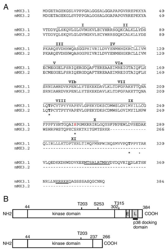
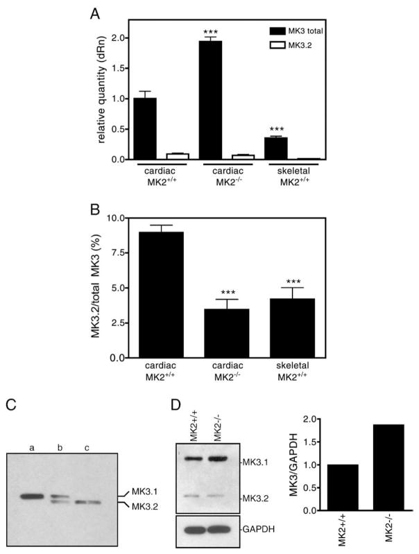
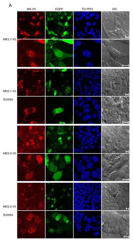
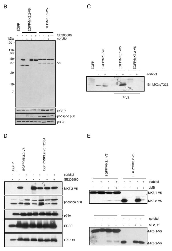
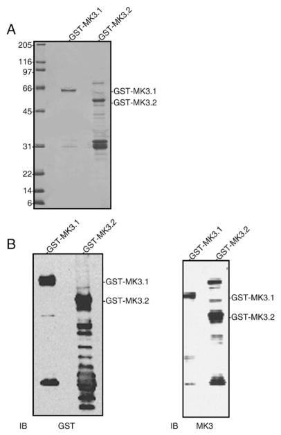
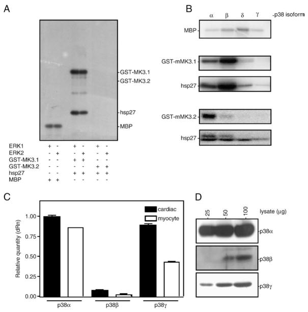
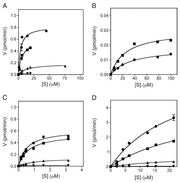
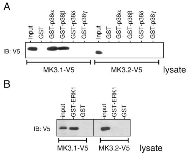
p38 MAP kinase (MAPK) isoforms alpha, beta, and gamma, are expressed in the heart. p38alpha appears pro-apoptotic whereas p38beta is pro-hypertrophic. The mechanisms mediating these divergent effects are unknown; hence elucidating the downstream signaling of p38 should further our understanding. Downstream effectors include MAPK-activated protein kinase (MK)-3, which is expressed in many tissues including skeletal muscles and heart. We cloned full-length MK3 (MK3.1, 384 aa) and a novel splice variant (MK3.2, 266 aa) from murine heart. For MK3.2, skipping of exons 8 and 9 resulted in a frame-shift in translation of the first 85 base pairs of exon 10 followed by an in-frame stop codon. Of 3 putative phosphorylation sites for p38 MAPK, only Thr-203 remained functional in MK3.2. In addition, MK3.2 lacked nuclear localization and export signals. Quantitative real-time PCR confirmed the presence of these mRNA species in heart and skeletal muscle; however, the relative abundance of MK3.2 differed. Furthermore, whereas total MK3 mRNA was increased, the relative abundance of MK3.2 mRNA decreased in MK2(-/-) mice. Immunoblotting revealed 2 bands of MK3 immunoreactivity in ventricular lysates. Ectopically expressed MK3.1 localized to the nucleus whereas MK3.2 was distributed throughout the cell; however, whereas MK3.1 translocated to the cytoplasm in response to osmotic stress, MK3.2 was degraded. The p38alpha/beta inhibitor SB203580 prevented the degradation of MK3.2. Furthermore, replacing Thr-203 with alanine prevented the loss of MK3.2 following osmotic stress, as did pretreatment with the proteosome inhibitor MG132. In vitro, GST-MK3.1 was strongly phosphorylated by p38alpha and p38beta, but a poor substrate for p38delta and p38gamma. GST-MK3.2 was poorly phosphorylated by p38alpha and p38beta and not phosphorylated by p38delta and p38gamma. Hence, differential regulation of MKs may, in part, explain diverse downstream effects mediated by p38 signaling.


Figures

Fig. 1
Fig. 1
cDNA and amino acid alignment of MK3 splice variants. (A) Predicted amino acid sequence of MK3.1 and MK3.2. Putative sites of phosphorylation by p38 MAPK (Thr-203, Ser-253, and Thr-315) are indicated by an asterisk and the auto-phosphorylation site (Thr-319) is indicated by a dot. Roman numerals above refer to the subdomains (I–IX) conserved in all protein kinases. The consensus bipartite nuclear localization signal is underlined whereas the putative nuclear export sequence is indicated by a double underline. Dashes represent gaps. (B) Schematic representation of MK3.1 and MK3.2. ‘E’ nuclear export signal; ‘L’, nuclear localization signal.
Fig. 2
Fig. 2
Detection of the MK3.1 and MK3.2 in murine ventricular myocardium. (A) The relative expression levels of total MK3 and MK3.2 mRNA were measured in RNA isolated from murine ventricular myocardium (filled bars) or skeletal muscle (open bars) of wild-type (MK2+/+) or MK2−/− mice by qPCR. Values are normalized to the amount of total MK3 transcript in hearts from MK2+/+ litter mates. (B) Abundance of MK3.2 mRNA expressed as percentage of total MK3 mRNA. (C) MK3.1 and MK3.2 protein are detected in mouse heart by immunoblotting. Recombinant GST-MK3.1 (lane a) and GST-MK3.2 (lane c), treated with thrombin to remove the GST moiety, were electrophoresed alongside 100 μg of lysate prepared from mouse ventricular myocardium (lane b). (D) Detection of MK3 protein in lysates from MK2−/− hearts and MK2+/+ litter mates by immunoblotting. Shown on the right, immunoreactive bands were quantified and normalized to GAPDH content. qPCR data are the mean and S.E. (n=8–10). ***, p<0.001 vs. total MK3 in MK2+/+ hearts (A) or MK3.2/total MK3 in MK2+/+ hearts (B); one-way ANOVA with Newman–Keuls post-hoc analysis.
Fig. 3
Fig. 3
Characterization of recombinant, expressed MK3.1-V5 and MK3.2-V5. (A) Subcellular localization of MK3.1-V5 or MK3.2-V5 in response to osmotic stress. HEK293t cells were transfected with pIRES-MK3.1-V5-EGFP or pIRES-MK3.2-V5-EGFP, starved, stimulated for 15 min with 0.3 M of sorbitol, fixed, labeled with V5 antisera and visualized by confocal fluorescence microscopy. Scale bar is 10 μM. (B) Osmotic stress induces a rapid breakdown of MK3.2-V5. HEK293t cells were transfected with pIRES-EGFP, pIRES-MK3.1-V5-EGFP or pIRES-MK3.2-V5-EGFP and, where indicated, were starved, stimulated for 15 min with 0.3Mof sorbitol, rinsed with cold TBS, lysed, and V5 immunoreactivity revealed following SDS-PAGE and immunoblotting. Where indicated, cells were pretreated with SB203580 (10 μM) for 10 min and then maintained for an additional 15 min in the presence of SB203580 and in the presence or absence of 0.3M sorbitol. Membranes were then stripped using 0.2 M NaOH (2×10 min) and reprobed using EGFP-specific antisera. In separate immunoblots, employing the same samples, filters were probed for phosphorylated p38 MAPK, stripped, and reprobed for total p38α immunoreactivity. Numbers at the left indicate the positions of the molecular mass marker proteins (in kDa). (C) Osmotic stress induces the phosphorylation of MK3. HEK293t cells were transfected with pIRES-EGFP, pIRES-MK2-V5-EGFP, pIRES-MK3.1-V5-EGFP or pIRES-MK3.2-V5-EGFP and, where indicated, were starved, stimulated for 15 min with 0.3 Mof sorbitol, rinsed with cold TBS, lysed, and probed with an antibody against human MK2phospho-threonine-222 following SDS-PAGE and immunoblotting. MK3.1-V5 and MK3.2-V5 were immunoprecipitated from lysates (1 mg) using an anti-V5 antibody. (D) MK3.2-V5T203A does not degrade in response to osmotic stress. HEK293t cells were transfected with pIRES-EGFP, pIRES-MK3.2-V5-EGFP or pIRES-MK3.2-V5 T203A-EGFP and, where indicated, were treated as described for panel B. (E) Effect of leptomycin B and MG132 upon the stability of MK3.2 during osmotic stress. HEK293t cells were transfected with pIRES-MK3.1-V5-EGFP or pIRES-MK3.2-V5-EGFP and, where indicated, were starved, stimulated for 15 min with 0.3 M of sorbitol, rinsed with cold TBS, lysed, and V5 immunoreactivity revealed following SDS-PAGE and immunoblotting. Where indicated, cells were pretreated with leptomycin B (LMB; 20 nM, 30 min) [21] or MG132 (50 μM, 4 h) [59] and then maintained for an additional 15 min in the presence of LMB or MG132 and in the presence or absence of 0.3 M sorbitol. Cells were then rinsed with cold TBS, lysed, and V5 immunoreactivity revealed following SDS-PAGE and immunoblotting.
Fig. 3
Fig. 3
Characterization of recombinant, expressed MK3.1-V5 and MK3.2-V5. (A) Subcellular localization of MK3.1-V5 or MK3.2-V5 in response to osmotic stress. HEK293t cells were transfected with pIRES-MK3.1-V5-EGFP or pIRES-MK3.2-V5-EGFP, starved, stimulated for 15 min with 0.3 M of sorbitol, fixed, labeled with V5 antisera and visualized by confocal fluorescence microscopy. Scale bar is 10 μM. (B) Osmotic stress induces a rapid breakdown of MK3.2-V5. HEK293t cells were transfected with pIRES-EGFP, pIRES-MK3.1-V5-EGFP or pIRES-MK3.2-V5-EGFP and, where indicated, were starved, stimulated for 15 min with 0.3Mof sorbitol, rinsed with cold TBS, lysed, and V5 immunoreactivity revealed following SDS-PAGE and immunoblotting. Where indicated, cells were pretreated with SB203580 (10 μM) for 10 min and then maintained for an additional 15 min in the presence of SB203580 and in the presence or absence of 0.3M sorbitol. Membranes were then stripped using 0.2 M NaOH (2×10 min) and reprobed using EGFP-specific antisera. In separate immunoblots, employing the same samples, filters were probed for phosphorylated p38 MAPK, stripped, and reprobed for total p38α immunoreactivity. Numbers at the left indicate the positions of the molecular mass marker proteins (in kDa). (C) Osmotic stress induces the phosphorylation of MK3. HEK293t cells were transfected with pIRES-EGFP, pIRES-MK2-V5-EGFP, pIRES-MK3.1-V5-EGFP or pIRES-MK3.2-V5-EGFP and, where indicated, were starved, stimulated for 15 min with 0.3 Mof sorbitol, rinsed with cold TBS, lysed, and probed with an antibody against human MK2phospho-threonine-222 following SDS-PAGE and immunoblotting. MK3.1-V5 and MK3.2-V5 were immunoprecipitated from lysates (1 mg) using an anti-V5 antibody. (D) MK3.2-V5T203A does not degrade in response to osmotic stress. HEK293t cells were transfected with pIRES-EGFP, pIRES-MK3.2-V5-EGFP or pIRES-MK3.2-V5 T203A-EGFP and, where indicated, were treated as described for panel B. (E) Effect of leptomycin B and MG132 upon the stability of MK3.2 during osmotic stress. HEK293t cells were transfected with pIRES-MK3.1-V5-EGFP or pIRES-MK3.2-V5-EGFP and, where indicated, were starved, stimulated for 15 min with 0.3 M of sorbitol, rinsed with cold TBS, lysed, and V5 immunoreactivity revealed following SDS-PAGE and immunoblotting. Where indicated, cells were pretreated with leptomycin B (LMB; 20 nM, 30 min) [21] or MG132 (50 μM, 4 h) [59] and then maintained for an additional 15 min in the presence of LMB or MG132 and in the presence or absence of 0.3 M sorbitol. Cells were then rinsed with cold TBS, lysed, and V5 immunoreactivity revealed following SDS-PAGE and immunoblotting.
Fig. 4
Fig. 4
Expression of recombinant MK3 splice variants. (A) Aliquots of purified GST-MK3.1 and GST-MK3.2 were resolved by 10–20% acrylamide-gradient SDS-PAGE then stained with Coomassie blue. (B) Purified GST-MK3.1 and GST-MK3.2 were separated by 10–20% acrylamide-gradient SDS-PAGE, transferred to nitrocellulose and probed with antisera raised against GST (left) or MK3 (right). Numbers at the left indicate the positions of the molecular mass marker proteins (in kDa).
Fig. 5
Fig. 5
Phosphorylation of MK3 splice variants by ERK1/2 and p38 MAPK isoforms. Purified proteins GST-MK3.1 and GST-MK3.2 were incubated for 1 h at 30 °C with phosphorylated, activated forms of (A) ERK1, ERK2 or (B) each p38 MAPK isoform (5 mU). The specific activity of each MAPK was determined using myelin basic protein (5 μg of MBP). Following phosphorylation, samples were solubilized using SDS-PAGE sample buffer and separated using SDS-PAGE. Gels were dried and 32P incorporation determined by autoradiography. (C) The relative expression levels of p38α, p38β, and p38γ mRNA were measured in RNA isolated from murine cardiac ventricular myocardium (filled bars) or cardiac ventricular myocytes (open bars) of wild-type mice by qPCR using the primers listed in Supplemental Table 2. Quantitation by qPCR was performed in triplicate: the results shown are the means (±S.E.) of 3 (heart) or 2 (myocyte) biological replicates. (D) The expression of p38α, p38β, and p38γ proteins were examined in the indicated amounts of murine ventricular lysate by immunoblotting using isoform-specific antisera.
Fig. 6
Fig. 6
Kinetic analysis of p38 MAPK-mediated phosphorylation of MK2, MK3 and MK5. Different amounts of purified GST-MK3.1 (A), GST-MK3.2 (B), GST-MK2 (C), or GST-MK5 (D) were incubated with the phosphorylated, activated forms of p38α (■), p38β (●), p38δ (▲), or p38γ (▼). A time course of 32P incorporation was performed at 30 °C for each indicated [S] as described in Materials and methods. Following phosphorylation, samples were solubilized using SDS-PAGE sample buffer and separated using SDS-PAGE. Gels were stained with Coomassie Brilliant Blue R-250, dried, the substrate band excised, and 32P incorporation determined by liquid scintillation counting. The specific activity of the [γ32P]ATP was determined for each experiment.
Fig. 7
Fig. 7
MK3.2 does not bind to p38 MAPK or ERK1. To determine if there are direct interactions between MK3.2 and p38 (A) or ERK1 (B), lysates from HEK cells transfected with either MK3.1-V5 or MK3.2-V5 (1 mg) were incubated with recombinant GSTp38α, GST-p38β, GST-p38δ, GST-p38γ, GST-ERK1 (1 μg) or GST. Co-precipitated MK3.1-V5 or MK3.2-V5 was detected by immunoblotting using anti-V5 antisera.

Similar articles

Cited by

References

    1. Ludwig S, Engel K, Hoffmeyer A, Sithanandam G, Neufeld B, Palm D, Gaestel M, Rapp UR. Mol Cell Biol. 1996;16(12):6687. - PMC - PubMed
    1. Tibbles LA, Woodgett JR. Cell Mol Life Sci. 1999;55(10):1230. - PMC - PubMed
    1. Ammerer G. Curr Opin Genet Dev. 1994;4(1):90. - PubMed
    1. Cobb MH, Goldsmith EJ. J Biol Chem. 1995;270(25):14843. - PubMed
    1. Hanks SK, Quinn AM, Hunter T. Science. 1988;241:42. - PubMed

Publication types

MeSH terms

Substances