. 2010 Jul 15;70(14):5923-30.
doi: 10.1158/0008-5472.CAN-10-0655. Epub 2010 Jun 22.

Development of a lung cancer therapeutic based on the tumor suppressor microRNA-34

Affiliations

Development of a lung cancer therapeutic based on the tumor suppressor microRNA-34

Jason F Wiggins et al. Cancer Res. .

Abstract

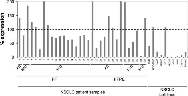
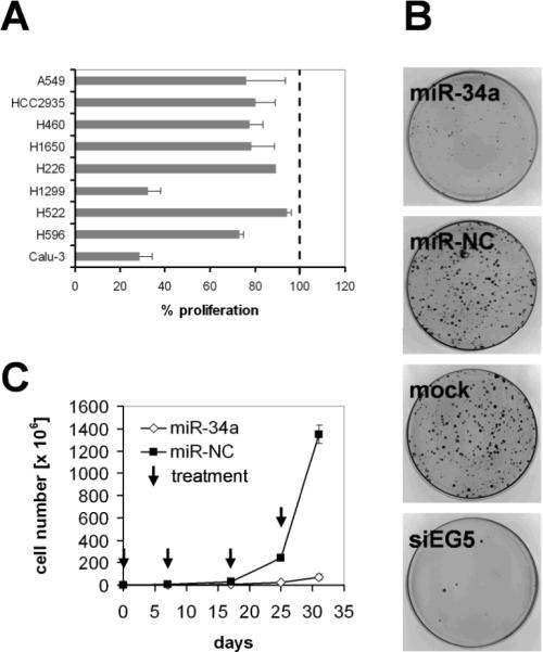
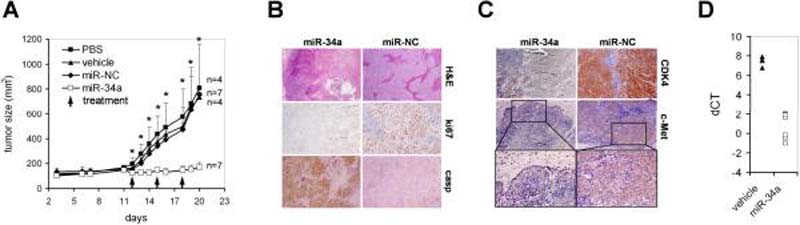
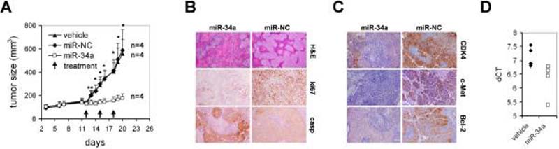
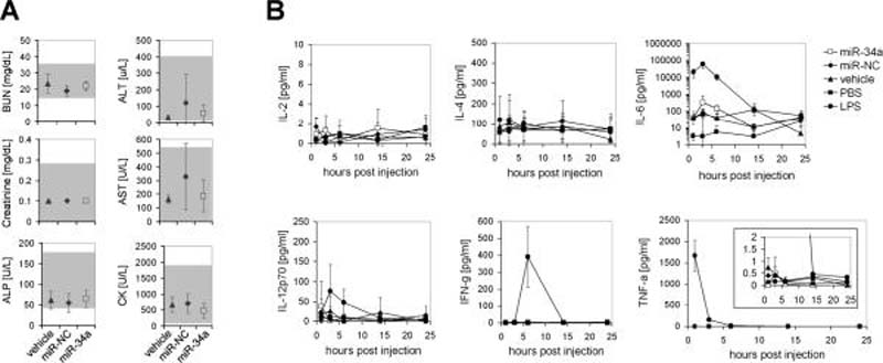
Tumor suppressor microRNAs (miRNA) provide a new opportunity to treat cancer. This approach, "miRNA replacement therapy," is based on the concept that the reintroduction of miRNAs depleted in cancer cells reactivates cellular pathways that drive a therapeutic response. Here, we describe the development of a therapeutic formulation using chemically synthesized miR-34a and a lipid-based delivery vehicle that blocks tumor growth in mouse models of non-small-cell lung cancer. This formulation is effective when administered locally or systemically. The antioncogenic effects are accompanied by an accumulation of miR-34a in the tumor tissue and downregulation of direct miR-34a targets. Intravenous delivery of formulated miR-34a does not induce an elevation of cytokines or liver and kidney enzymes in serum, suggesting that the formulation is well tolerated and does not induce an immune response. The data provide proof of concept for the systemic delivery of a synthetic tumor suppressor mimic, obviating obstacles associated with viral-based miRNA delivery and facilitating a rapid route for miRNA replacement therapy into the clinic.


Figures

Figure 1
Figure 1. miR-34a expression in human non-small cell lung cancer
qRT-PCR analysis of miR-34a using total RNA from 32 NSCLC tumor samples and their corresponding normal adjacent tissues (NAT), as well as total RNA from various NSCLC cell lines. Raw Ct values from lung tumors and NATs were normalized to a house-keeping miRNA (20) and expressed as % expression compared to levels in the respective NAT from the same patient (100%). Values derived from cell lines were compared to the expression value in Wi-38 normal lung fibroblasts (100%). FF, flash-frozen; FFPE, formalin-fixed paraffin-embedded; AC, adenocarcinoma; BAC, bronchioalveolar carcinoma; LCC, large cell carcinoma; SCC, squamous cell carcinoma.
Figure 2
Figure 2. Inhibitory activity of miR-34a in cultured lung cancer cells
(A) Transient effect of synthetic miR-34a. Cancer cell lines were transfected in triplicate with 30 nM miR-34a or miR-NC. Proliferation was assessed 3–7 d post transfection using Alamar Blue™ (Invitrogen). Percent (%) change in proliferation was plotted relative to proliferation of miR-NC transfected cells. Averages and standard deviations of three replicates are shown. (B) Colony formation assay using SW-900 lung cancer cells. Cells were transfected with synthetic miR-34a, miR-NC and siEG5 siRNA and seeded at 3000 cells per 100-mm dish. After 32 days, cells were stained with 2% crystal violet. Colonies containing >50 cells were counted. (C) Long-term effects of miR-34a. H226 cells were transfected in triplicate with miR-34a and miR-NC, seeded and propagated in regular growth medium. When the control cells reached confluence, cells were harvested, counted and transfected again with the respective miRNAs. The population doublings were calculated, and cell counts were extrapolated and plotted on a linear scale. Arrows represent transfection events. Average cell counts and standard deviations of three replicates are shown. Cell lines carrying mutated p53 include Calu-3, H596, H1299, H522 and SW-900 (21, 22). A549 and H460 carry wild-type p53 (21, 22).
Figure 3
Figure 3. Local delivery of formulated miR-34a inhibits lung tumor growth in mice
(A) Effects of formulated miR-34a in H460 tumors by intratumoral injections are shown. Palpable subcutaneous H460 tumor xenografts were treated on days 12, 15 and 18 with each 100 μg formulated miR-34a or miR-NC. As controls, separate groups of tumor-bearing animals were injected with vehicle alone or phosphate-buffered saline (PBS). Caliper measurements were taken on days as indicated and averaged. Error bars indicate standard deviations. **, p values <0.05 (Student's t-test, 2-tailed, miR-34a and miR-NC); *, p values <0.01 (Student's t-test, 2-tailed, miR-34a and miR-NC). (B,C) Histologies and immunohistochemistry stainings directed against ki67, caspase 3, CDK4, c-Met of H460 tumors after sacrifice. Histologies at a 100-fold magnification and immunohistochemistry stainings at a ~300-fold magnification are shown. Insets show c-Met-specific stainings at a 400-fold magnification. (D) qRT-PCR analysis of miR-34a in H460 tumors treated with formulated miR-34a or vehicle after sacrifice. Raw Ct values from lung tumors and NATs were normalized to a house-keeping miRNA (20) and expressed as dCT [log2].
Figure 4
Figure 4. Intravenous delivery of formulated miR-34a blocks growth of human lung tumors in mice
(A) Mice carrying palpable subcutaneous H460 tumor xenografts were treated on days 12, 15 and 18 with lipid-formulated miR-34a, miR-NC, vehicle alone or phosphate-buffered saline (PBS) by intravenous tail vein injections. Each dose contained 100 μg of formulated oligo. Caliper measurements were taken on days as indicated and averaged. Error bars indicate standard deviations. **, p values <0.05 (Student's t-test, 2-tailed, miR-34a and miR-NC); *, p values <0.01 (Student's t-test, 2-tailed, miR-34a and miR-NC). (B,C) Histologies and immunohistochemistry stainings specific for ki67, caspase 3, CDK4, c-Met and Bcl-2 of H460 tumors after sacrifice. Histologies at a 100-fold magnification and immunohistochemistry stainings at a ~300-fold magnification are shown. (D) qRT-PCR analysis of miR-34a in H460 tumors treated with formulated miR-34a or vehicle after sacrifice. Raw Ct values from lung tumors and NATs were normalized to a house-keeping miRNA (20) and expressed as dCT [log2].
Figure 5
Figure 5. Blood chemistries and cytokine levels in response to systemic delivery of formulated miR-34a
(A) Serum levels of blood urea nitrogen (BUN), creatinine, alkaline phosphatase (ALP), alanine transaminase (ALT), aspartate transaminase (AST) and creatine kinase (CK) of animals shown in Fig. 4A after sacrifice. Averages and standard deviations of all 4 animals are shown. The gray-shaded area indicates guideline ranges as reported by the Comparative Pathology Laboratory at UC Davis, CA. (B) Serum cytokine levels in immunocompetent Balb/c mice 1–24 hrs post intravenous injection of a single dose of formulated miR-34a, miR-NC, vehicle and PBS. Lipopolysaccharide (LPS) was used as a positive control for IL-6, IFN- γ and TNF-α induction. Averages and standard deviations of 3 animals per group are shown. IL-6 data are shown on a logarithmic scale.

Similar articles

Cited by

References

    1. Bartel DP. MicroRNAs: genomics, biogenesis, mechanism, and function. Cell. 2004;116:281–97. - PubMed
    1. Esquela-Kerscher A, Slack FJ. Oncomirs - microRNAs with a role in cancer. Nat Rev Cancer. 2006;6:259–69. - PubMed
    1. Calin GA, Croce CM. MicroRNA signatures in human cancers. Nat Rev Cancer. 2006;6:857–66. - PubMed
    1. Petrocca F, Lieberman J. Micromanipulating cancer: microRNA-based therapeutics? RNA Biol. 2009;6:335–40. - PubMed
    1. Tong AW, Nemunaitis J. Modulation of miRNA activity in human cancer: a new paradigm for cancer gene therapy? Cancer Gene Ther. 2008;15:341–55. - PubMed

Publication types

MeSH terms