. 2010 Jul;137(14):2417-26.
doi: 10.1242/dev.047993.

The Adam family metalloprotease Kuzbanian regulates the cleavage of the roundabout receptor to control axon repulsion at the midline

Affiliations

The Adam family metalloprotease Kuzbanian regulates the cleavage of the roundabout receptor to control axon repulsion at the midline

Hope A Coleman et al. Development. 2010 Jul.

Abstract

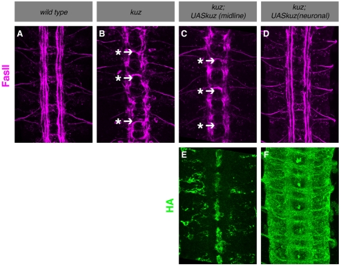
Slits and their Roundabout (Robo) receptors mediate repulsive axon guidance at the Drosophila ventral midline and in the vertebrate spinal cord. Slit is cleaved to produce fragments with distinct signaling properties. In a screen for genes involved in Slit-Robo repulsion, we have identified the Adam family metalloprotease Kuzbanian (Kuz). Kuz does not regulate midline repulsion through cleavage of Slit, nor is Slit cleavage essential for repulsion. Instead, Kuz acts in neurons to regulate repulsion and Kuz can cleave the Robo extracellular domain in Drosophila cells. Genetic rescue experiments using an uncleavable form of Robo show that this receptor does not maintain normal repellent activity. Finally, Kuz activity is required for Robo to recruit its downstream signaling partner, Son of sevenless (Sos). These observations support the model that Kuz-directed cleavage is important for Robo receptor activation.


Figures

Fig. 1.
Fig. 1.
Genetic interactions between kuz, slit, robo and comm. (A-L) Stage 16 embryos were stained with MAb BP102 (A-D,K,L) or MAb FasII (E-J) to reveal all CNS axons or subsets of ipsilateral axons, respectively. Anterior is up. (A) A wild-type embryo exhibiting the characteristic ladder-like axon scaffold. (B) A robo mutant embryo; note the reduction in thickness of the longitudinal connectives and the commensurate thickening of the commissures. (C) kuz mutants also show thinning of the longitudinals and thickening of the commissures. (D) In kuz mutants that are simultaneously heterozygous for slit and robo, the thickening of the commissures is qualitatively enhanced. (E) A wild-type embryo has three bundles of FasII-positive axons that do not cross the midline. (F) In robo mutant embryos, the medial bundle of FasII-positive axons wanders back and forth across the midline (arrowheads with asterisks). (G) kuz zygotic mutants have a milder crossing defect (arrows with asterisks), a phenotype that is enhanced when slit and robo are also heterozygous (H). (I) A slit, robo/+ embryo with a mild midline crossing defect (arrow with asterisk). (J) An example of a slit, robo/+, kuz/+ embryo showing dominant enhancement (arrows with asterisks). (K) comm mutant embryos completely lack axon commissures, whereas kuz; comm double mutants reveal a partial restoration of commissure formation (arrows in L).
Fig. 2.
Fig. 2.
Misexpression of UASkuzDN results in ectopic crossing of apterous neurons. (A-C) Stage 16 apGal4, UASTau-Myc-GFP embryos were stained with a polyclonal antibody against GFP. Anterior is up. In wild-type embryos (A) the ap axons do not cross the midline, whereas misexpression of UASKuzDN driven by apGal4 (B) results in ectopic crossing (arrows with asterisks). (C) Removing one copy of slit in embryos mis-expressing UASKuzDN enhances the crossing phenotype. (D) Quantification shows the percentage of segments in which the ap axons cross the midline. **P<0.0001, *P<0.05 (unpaired Student's t-test). Error bars indicate s.d.
Fig. 3.
Fig. 3.
kuz is required in neurons. (A-F) Stage 16 embryos were stained with a mAb to FasII (1D4). Anterior is up. (A) A wild-type embryo stained with mAb 1D4. (B) Ipsilateral axons from the medial and intermediate fascicles aberrantly cross the midline in kuz mutants (arrow with asterisk). (C,E) Midline expression of UASkuzHA using a slitGal4 driver does not rescue the kuz phenotype. One hundred percent of kuz mutant embryos with midline kuz overexpression show ectopic midline crossing of FasII-positive axons in greater than half of all segments (107 out of 135 or 79% of segments show ectopic crossing, n=15 embryos). (D,F) Pan-neural expression of UASkuzHA provides complete rescue of the kuz mutant phenotype (0 out of 144 segments show ectopic crossing, n=16 embryos).
Fig. 4.
Fig. 4.
Slit-U rescues the slit mutant phenotype. (A) Schematic drawing of Slit. The arrow indicates the cleavage site. Slit-U or Slit-FL were expressed in HEK293T cells. Total lysates of the cells were harvested and subjected to polyacrylamide gel electrophoresis (PAGE). (B,C) Western blotting with antibodies to either the N- or the C-terminus of Slit indicates that Slit-U is not processed in 293T cells (B) or in embryos (C). (D) Quantification of segments in which FasII-positive neurons cross the midline. Error bars indicate s.d. (E-H) Stage 16 embryos were stained with a mAb to FasII (1D4). Anterior is up. (E) A wild-type embryo stained with mAb 1D4. (F) A slit mutant embryo. All axons collapse on the midline and there are no longer any distinct fascicles (asterisk). Expression of UASslit-FL (G) or UASslit-U (H) driven by slitGal4 in a slit embryo mostly rescues the aberrant midline crossing phenotype, but does not restore proper lateral positioning of the FasII-positive fascicles (arrow with asterisk).
Fig. 5.
Fig. 5.
Kuz promotes Robo cleavage in vitro. UASroboGFP with or without UASkuzHA were transfected into Drosophila S2R+cells. (A) Western blotting of separated proteins harvested from the media with antibodies to a N-terminal epitope of Robo reveals a significant increase in the amount of Robo ectodomain in the media of cells co-transfected with UASkuzHA compared with that of cells with no ectopic expression of kuz (lanes 1, 4 and 5). Co-transfection of UASrobo and UASkuzDmetallo did not increase the amount of Robo ectodomain detected in the media (lanes 1, 2 and 3). A lane between the first and second lane shown was excised from the media blot. Western blotting of the total lysates with antibodies directed against GFP, HA and tubulin show the relative levels of RoboGFP, KuzHA or KuzΔmetallo, and tubulin, respectively. (B) Western blotting of protein harvested from the media of cells transfected with UASHAroboMyc with or without UASkuzHA using an antibody directed against HA. (C) Western blots of proteins harvested from dsRNA-treated S2 cells. Cells expressing UASroboGFP, UASkuzHA, or both were treated with kuz dsRNA. The level of Robo ectodomain detected in the media from cells expressing both robo and kuz is lower in cells treated with kuz dsRNA (lanes 3 and 4). Treatment with kuz dsRNA also reduces the amount of Robo ectodomain detected in the media of cells with no transfected kuz (lanes 1 and 2). Western blotting of the total lysates with antibodies directed against Robo, HA and tubulin show the relative levels of RoboGFP, KuzHA and tubulin, respectively.
Fig. 6.
Fig. 6.
Surface levels of Robo are increased and mislocalized in kuz mutants. (A-F) Stage 15 kuz/+ heteroygotes (A-C) or kuz mutant (D-F) embryos were dissected and stained live to reveal Robo surface expression. Anterior is up. Embryos were stained together on the same slide and micrographs were generated using identical confocal settings. (A-C) A kuz/+ heterozygous embryo stained with anti-HRP to reveal all axons (A) and with anti-Robo to visualize Robo distribution (B). Robo is enriched on longitudinal connectives and almost absent from axon commissures (compare B and C). (D-F) A kuz mutant embryo exhibits increased levels of Robo expression (compare E and B) and Robo protein is no longer restricted from the axon commissures (E,F). (G) A western blot from embryonic extracts of the indicated genotypes reveals increased Robo expression levels. Anti-tubulin was used as a loading control. (H) Quantification of Robo fluorescence intensity reveals a significant increase in Robo surface expression in kuz mutants. Each bar in the histogram represents an individual embryo. Fluorescence intensity was calculated as previously reported (Yang and Bashaw, 2006).
Fig. 7.
Fig. 7.
Expression of an uncleavable form of Robo does not rescue robo mutants. (A) Schematic depicting the receptors used in the rescue experiment. In Robo-U the Fn domains of Robo are swapped for the first three Fn domains of Fra. The Fra-Ro receptor has the ectodomain of Fra and the transmembrane and intracellular domains of Robo. (B) The media and total lysates of S2 cells expressing the receptors depicted in A, with or without added UASkuzHA, were subjected to PAGE and subsequent western blotting as previously described. No ectodomain shedding was detected from cells expressing Fra-RoMyc, Robo-UMyc, or FraMyc. Western blotting of the total lysates with antibodies directed against Myc, HA and tubulin show the relative levels of Myc-tagged receptor, KuzHA and tubulin, respectively. (C) Quantification of the number of segments in which ap axons ectopically cross the midline in robo mutants expressing different forms of the Robo receptor under the control of apGal4. Shown is the percentage of segments in which the ap axons cross the midline. *P<0.0001 (unpaired Student's t-test) compared to ap axons expressing wild-type Robo. Error bars indicate s.d. (D-H) Stage 16 embryos were stained with a MAb to FasII (1D4). Anterior is up. (D) A wild-type embryo stained with mAb 1D4. (E) In robo mutants, the medial-most fascicles cross the midline (arrows with asterisks). In robo embryos expressing Robo-UMyc (F) or Fra-RoMyc (G) under the control of the elavGal4 driver, the medial-most fascicles still cross the midline in many segments (arrows with asterisks). (H) Pan-neural (elavGal4) expression of wild-type Robo provides significant rescue of the robo phenotype, although one can still observe infrequent ectopic crossing of FasII-positive axons (arrow with asterisk). The small panels below the FasII panels indicate the expression levels and patterns of the Myc-tagged receptors. Arrowheads indicate the midline; asterisks indicate Myc-tagged receptor expression on crossing axons. (I-M) Stage 16 embryos were stained with a polyclonal antibody directed against GFP to detect the ap neurons. Anterior is up. (I) In wild-type embryos the ap neurons never cross the midline. (J) In robo mutants, the ap neurons collapse upon the midline. In robo mutants expressing Robo-U (K) or Fra-Ro (L) driven by apGal4, the ap axons from either side of the midline are fused and collapsed on the midline in some segments (<*), but are correctly routed on either side of the midline in others (<). (M) Expression of wild-type Robo in ap neurons in a robo mutant background rescues the ectopic crossing phenotype.
Fig. 8.
Fig. 8.
Expression of mouse KuzDN blocks Slit-dependent recruitment of Sos to the plasma membrane. HEK293T cells were transiently transfected with human Myc-His-ROBO1. Endogenous human SOS is visualized by rabbit anti-mSos1 antibodies (green), human ROBO1 is visualized by mouse anti-Myc antibodies (blue), and F-actin is visualized by rhodamine-conjugated phalloidin (magenta). A single confocal section is shown. (A-D) A ROBO1-expressing cell treated with control medium. The morphology of the cell is elongated and SOS is predominantly localized in the cytoplasm. (E-H) A ROBO1-expressing cell treated with SLIT2-conditioned medium for five minutes. The cell morphology has become remarkably rounded and SOS proteins are recruited to the membrane, partially colocalizing with ROBO1 (white in H). (I-L) Cells expressing both ROBO1 and mouse KuzDN treated with SLIT2 medium. The cells retain their elongated morphology and endogenous SOS localization is cytosolic. (M) Quantification of the ratio of the average pixel density of SOS staining colocalized with the membrane to the average pixel density of cytosolic SOS. Shown is the average of the ratio of membrane to cytosolic SOS. *P<0.0001 (unpaired Student's t-test) compared with ROBO1-expressing cells treated with bath application of SLIT2. Error bars indicate s.d.
Fig. 9.
Fig. 9.
Model of Kuz function in Slit/Robo repulsion. Our data suggest a model for Kuz function in the Slit/Robo pathway in which Slit binding to the Robo receptor results in cleavage of Robo by Kuz. Release of the Robo ectodomain could cause a conformational change in the remainder of the membrane-bound Robo receptor that strengthens its interaction with the Dock and recruits Sos to the plasma membrane. Sos is then properly localized to the plasma membrane, where it activates Rac to promote growth cone repulsion.

References

    1. Bashaw G. J., Goodman C. S. (1999). Chimeric axon guidance receptors: the cytoplasmic domains of slit and netrin receptors specify attraction versus repulsion. Cell 97, 917-926 - PubMed
    1. Battye R., Stevens A., Jacobs J. R. (1999). Axon repulsion from the midline of the Drosophila CNS requires slit function. Development 126, 2475-2481 - PubMed
    1. Becherer J. D., Blobel C. P. (2003). Biochemical properties and functions of membrane-anchored metalloprotease-disintegrin proteins (ADAMs). Curr. Top. Dev. Biol. 54, 101-123 - PubMed
    1. Beel A. J., Sanders C. R. (2008). Substrate specificity of gamma-secretase and other intramembrane proteases. Cell. Mol. Life Sci. 65, 1311-1334 - PMC - PubMed
    1. Brand A. H., Perrimon N. (1993). Targeted gene expression as a means of altering cell fates and generating dominant phenotypes. Development 118, 401-415 - PubMed

Publication types

Substances