Analysis of X chromosome genomic DNA sequence copy number variation associated with premature ovarian failure (POF)
- PMID: 20570974
- PMCID: PMC3836253
- DOI: 10.1093/humrep/deq158
Analysis of X chromosome genomic DNA sequence copy number variation associated with premature ovarian failure (POF)
Abstract
Background: Premature ovarian failure (POF) is a heterogeneous disease defined as amenorrhoea for >6 months before age 40, with an FSH serum level >40 mIU/ml (menopausal levels). While there is a strong genetic association with POF, familial studies have also indicated that idiopathic POF may also be genetically linked. Conventional cytogenetic analyses have identified regions of the X chromosome that are strongly associated with ovarian function, as well as several POF candidate genes. Cryptic chromosome abnormalities that have been missed might be detected by array comparative genomic hybridization.
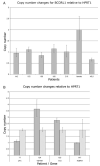
Methods: In this study, samples from 42 idiopathic POF patients were subjected to a complete end-to-end X/Y chromosome tiling path array to achieve a detailed copy number variation (CNV) analysis of X chromosome involvement in POF. The arrays also contained a 1 Mb autosomal tiling path as a reference control. Quantitative PCR for selected genes contained within the CNVs was used to confirm the majority of the changes detected. The expression pattern of some of these genes in human tissue RNA was examined by reverse transcription (RT)-PCR.
Results: A number of CNVs were identified on both Xp and Xq, with several being shared among the POF cases. Some CNVs fall within known polymorphic CNV regions, and others span previously identified POF candidate regions and genes.
Conclusions: The new data reported in this study reveal further discrete X chromosome intervals not previously associated with the disease and therefore implicate new clusters of candidate genes. Further studies will be required to elucidate their involvement in POF.
Figures


Comment in
-
Array comparative genomic hybridization for the detection of submicroscopic copy number variations of the X chromosome in women with premature ovarian failure.Hum Reprod. 2010 Dec;25(12):3159-60; author reply 3160-1. doi: 10.1093/humrep/deq284. Epub 2010 Oct 16. Hum Reprod. 2010. PMID: 20952765 No abstract available.
References
-
- Aboura A, Dupas C, Tachdjian G, Portnoï MF, Bourcigaux N, Dewailly D, Frydman R, Fauser B, Ronci-Chaix N, Donadille B, et al. Array comparative genomic hybridization profiling analysis reveals deoxyribonucleic acid copy number variations associated with premature ovarian failure. J Clin Endocrinol Metab. 2009;94:4540–4546. - PubMed
-
- Bertini V, Ghirri P, Bicocchi MP, Simi P, Valetto A. Molecular cytogenetic definition of a translocation t(X;15) associated with premature ovarian failure. Fertil Steril. 2010 Mar 23; [Epub ahead of print] - PubMed
-
- Bione S, Rizzolio F, Sala C, Ricotti R, Goegan M, Manzini MC, Battaglia R, Marozzi A, Vegetti W, Dalprà L, et al. Mutation analysis of two candidate genes for premature ovarian failure, DACH2 and POF1B. Hum Reprod. 2004;19:2759–2766. - PubMed
-
- Bodega B, Bione S, Dalpra L, Toniolo D, Ornaghi F, Vegetti W, Ginelli E, Marozzi A. Influence of intermediate and uninterrupted FMR1 CGG expansions in premature ovarian failure manifestation. Hum Reprod. 2006;21:952–957. - PubMed
Publication types
MeSH terms
Grants and funding
LinkOut - more resources
Full Text Sources
Other Literature Sources
Medical
Miscellaneous

