Review
. 2010 Jul;6(7):380-95.
doi: 10.1038/nrendo.2010.71.

A local autocrine axis in the testes that regulates spermatogenesis

Affiliations
Review

A local autocrine axis in the testes that regulates spermatogenesis

C Yan Cheng et al. Nat Rev Endocrinol. 2010 Jul.

Abstract

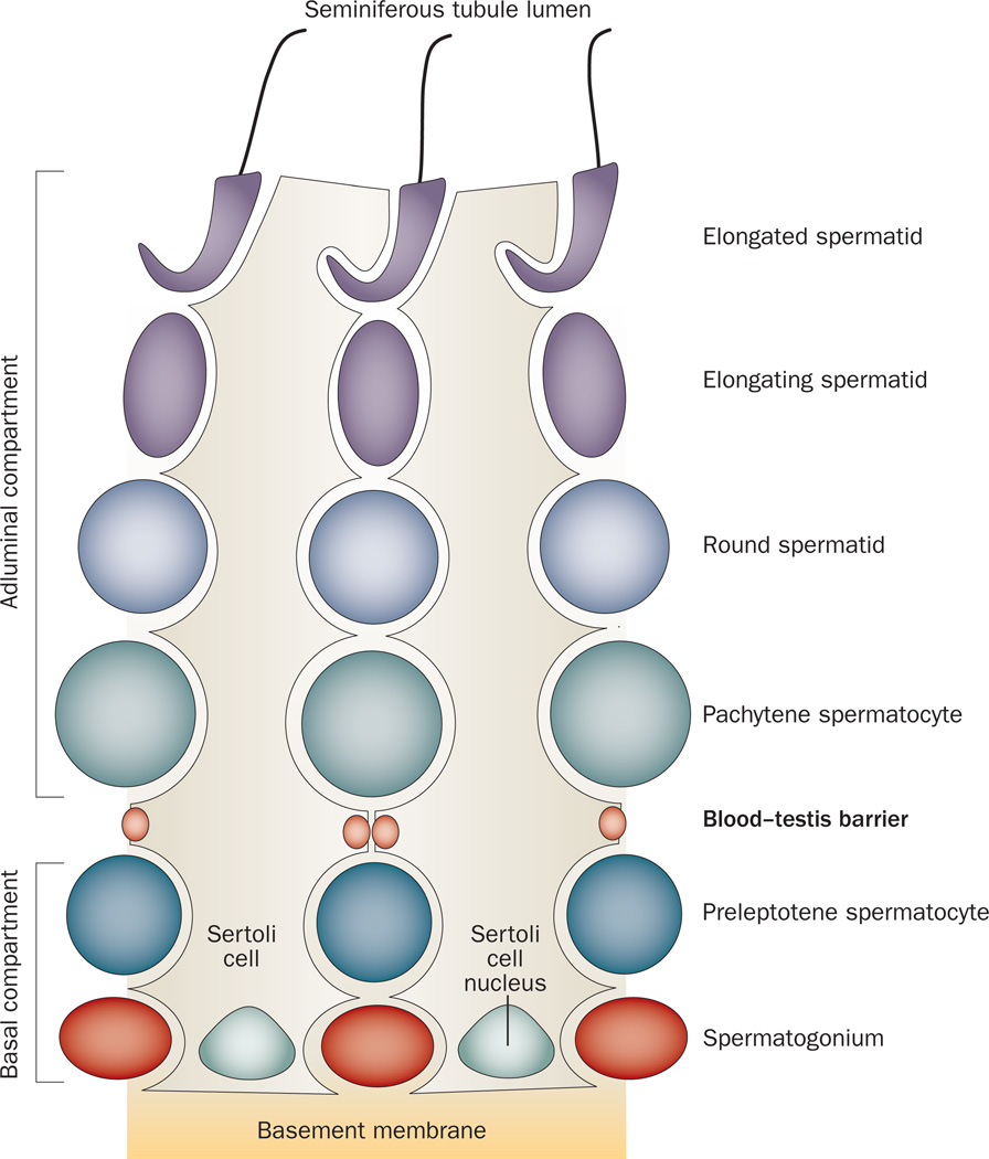
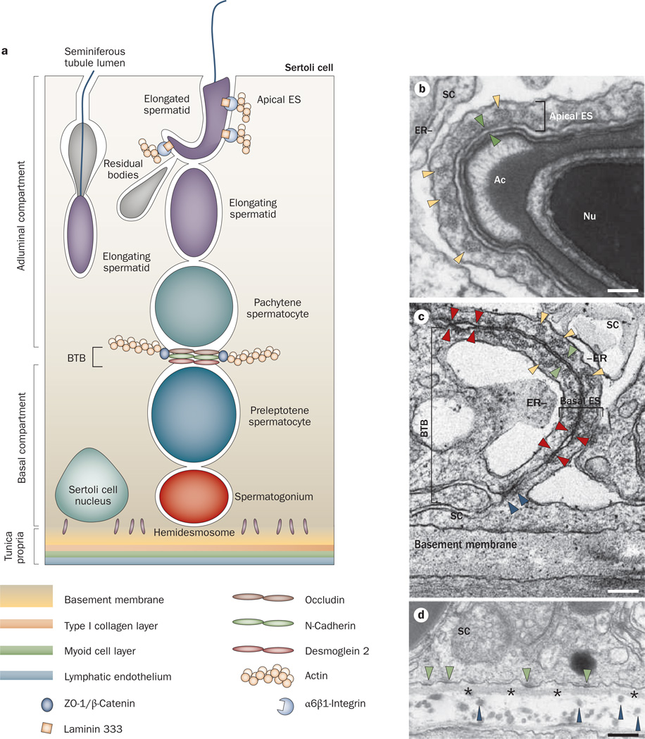
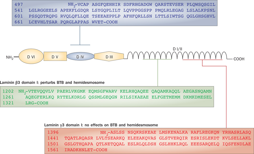
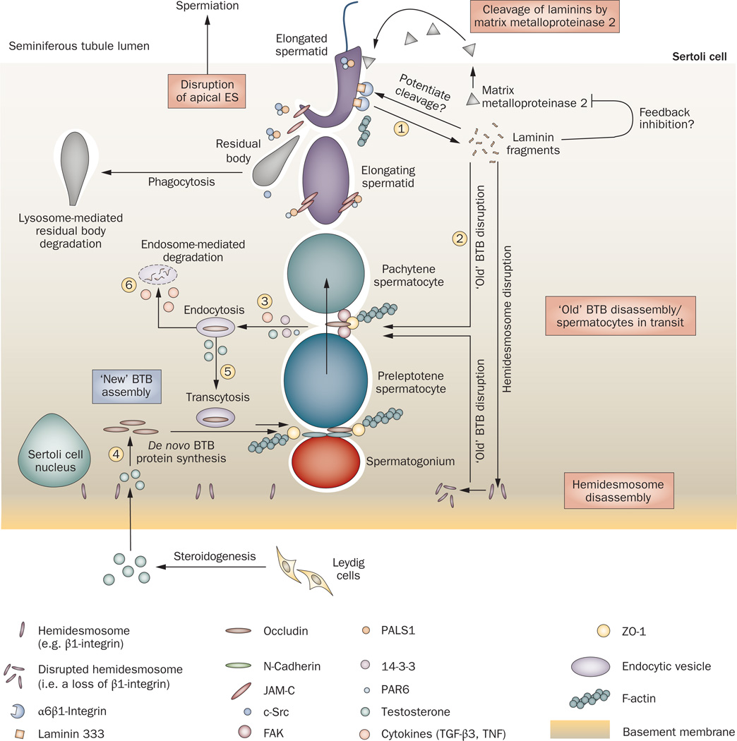
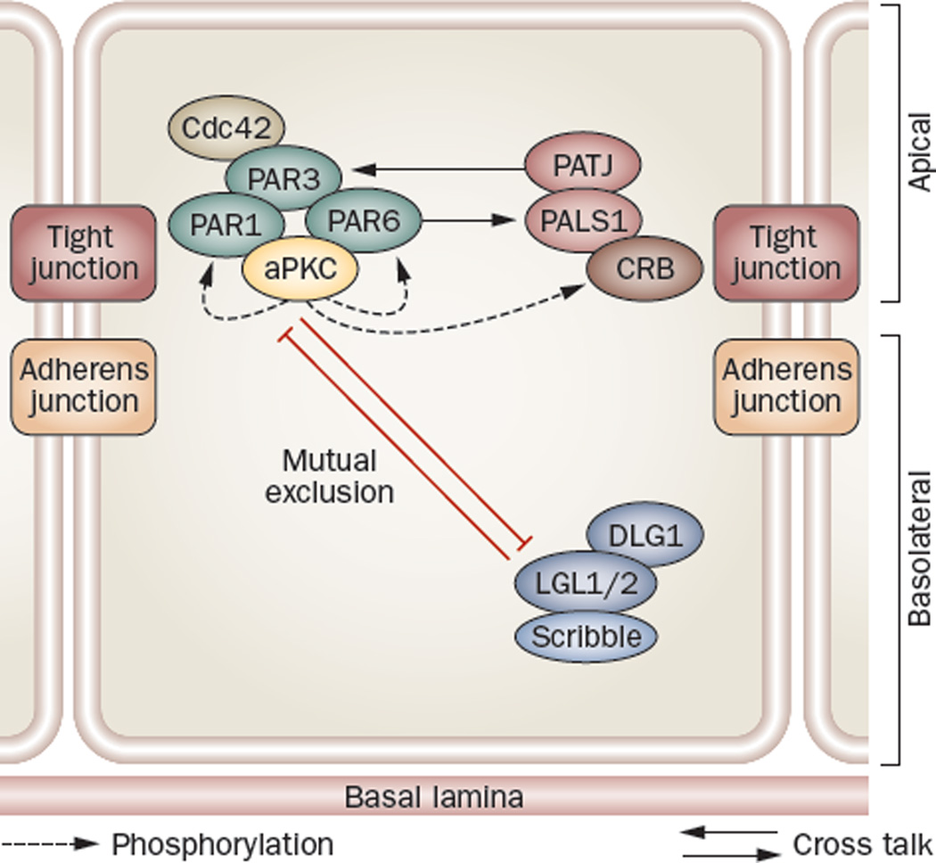
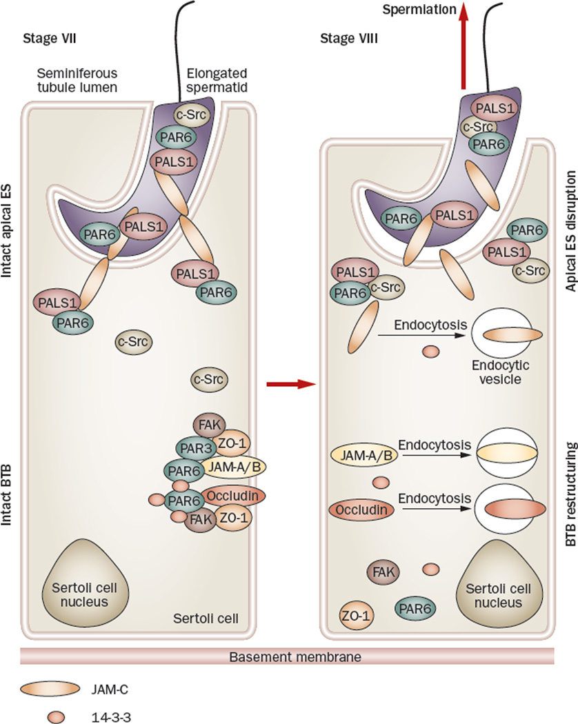
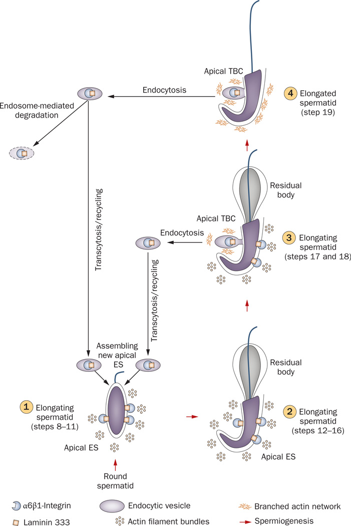
Spermiation--the release of mature spermatozoa from Sertoli cells into the seminiferous tubule lumen--occurs by the disruption of an anchoring device known as the apical ectoplasmic specialization (apical ES). At the same time, the blood-testis barrier (BTB) undergoes extensive restructuring to facilitate the transit of preleptotene spermatocytes. While these two cellular events take place at opposite ends of the Sertoli cell epithelium, the events are in fact tightly coordinated, as any disruption in either process will lead to infertility. A local regulatory axis exists between the apical ES and the BTB in which biologically active laminin fragments produced at the apical ES by the action of matrix metalloproteinase 2 can regulate BTB restructuring directly or indirectly via the hemidesmosome. Equally important, polarity proteins play a crucial part in coordinating cellular events within this apical ES-BTB-hemidesmosome axis. Additionally, testosterone and cytokines work in concert to facilitate BTB restructuring, which enables the transit of spermatocytes while maintaining immunological barrier function. Herein, we will discuss this important autocrine-based cellular axis that parallels the hormonal-based hypothalamic-pituitary-testicular axis that regulates spermatogenesis. This local regulatory axis is the emerging target for male contraception.


Figures

Figure 1
Figure 1
Cross-section of a mammalian seminiferous tubule, which is composed of the seminiferous epithelium constituted by Sertoli cells and germ cells at different stages of their development. During spermiation, fully developed spermatids detach from the seminiferous epithelium, enter the tubule lumen and are transported to the epididymis for maturation. if an investigator is viewing a specific section of a seminiferous tubule under a stereomicroscope, the unique association of developing spermatids (round spermatids, elongating spermatids, and elongated spermatids) in the seminiferous epithelium relative to Sertoli cells and other germ cells, namely spermatogonia and spermatocytes, occur in cycles (or waves), and these cycles are classified into stages, with each stage composed of some unique cellular events pertinent to spermatogenesis. For instance, in adult rats, at stage VIII of the epithelial cycle, spermiation takes place simultaneously with the transit of preleptotene spermatocytes at the blood–testis barrier, and at stage XIV, meiosis I and II occur.
Figure 2
Figure 2
Anatomy of the seminiferous epithelium in the mammalian testis. a | Anatomical features and the spatial relationship of the apical ES, the BTB and the hemidesmosome in adult rat testes. The BTB with coexisting tight junctions (occludin), basal ES (N-cadherin), and desmosome–gap junctions (desmoglein 2) physically divides the seminiferous epithelium into basal and apical (adluminal) compartments. The apical ES is constituted by different protein complexes, such as α6β1-integrin–laminin 333. b | The apical ES at the elongated spermatid–Sertoli cell interface is typified by the presence of actin filament bundles (yellow arrowheads), which are sandwiched in between cisternae of ER and the apposing plasma membrane (see apposing green arrowheads) of the spermatid and the Sertoli cell. These ultrastructures are restricted to the Sertoli cell. c | The BTB is present at the base of two adjacent Sertoli cells near the basement membrane. it is constituted by coexisting tight junctions (denoted by apposing red arrowheads), basal ES and desmosome–gap junctions (noted by the electron-dense material between two adjacent Sertoli cells, see blue arrowheads). The basal ES is typified by the presence of actin filament bundles (yellow arrowheads) sandwiched in between cisternae of ER and the apposing Sertoli cell plasma membrane (see apposing green arrowheads), which is similar to the apical ES except that these ultrastructural features are present within both Sertoli cells. d | The hemidesmosome is found at the interface between the Sertoli cell and the basement membrane (asterisks) typified by patches of electron-dense material (see green arrowheads). Also noted are type I collagen fibrils (blue arrowheads) found below the basement membrane. Bar in panel b, 0.1 µm; panel c, 0.12 µm; panel d, 0.15 µm. Abbreviations: Ac, acrosome; BTB, blood–testis barrier; ER, endoplasmic reticulum; ES, ectoplasmic specialization; Nu, spermatid nucleus; SC, Sertoli cell.
Figure 3
Figure 3
Structural domains of a typical laminin chain. Laminin β3 and γ3 are found at the apical ES in the rat testis. Three laminin fragments have been examined. Laminin β3 domain I and γ3 domain IV, but not laminin γ3 domain I, were shown to be biologically active fragments that affected Sertoli cell BTB dynamics and hemidesmosome function. Abbreviation: BTB, blood–testis barrier.
Figure 4
Figure 4
The apical ES–BTB–hemidesmosome functional axis in the testis. The cascade of events begins at the apical ES before spermiation. Matrix metalloproteinase 2, which is expressed predominantly before spermiation, cleaves the integrin–laminin protein complex at the apical ES to facilitate spermiation, generating the biologically active laminin fragments (1). Whether biologically active laminin fragments induce further proteolytic events to disrupt the apical ES locally and/or a feedback mechanism prevents further damage to the epithelium resulting from matrix metalloproteinase 2 activation is not known. Once biologically active laminin fragments are formed, they induce disruption of tight junction fibrils at the ‘old’ BTB site above preleptotene spermatocytes in transit across the BTB (2) and also disrupt the hemidesmosome. This loss of hemidesmosome function, as manifested by a decline in β1-integrin at the hemidesmosome, potentiates disruption at the ‘old’ BTB site to facilitate the transit of spermatocytes, which is mediated by an increase in protein endocytosis at the site, (3); this endocytosis is facilitated by cytokines (TGF-β3 and TNF),, testosterone, PAR polarity proteins (14-3-3 and PAR6), and kinases (such as FAK and c-Src)., To maintain the immunological barrier during the transit of primary spermatocytes, ‘new’ tight junction fibrils are probably first formed behind migrating spermatocytes, mediated by the de novo synthesis of BTB integral membrane proteins induced by testosterone–, (4) possibly via both classical and nongenomic pathways, so that a ‘new’ BTB can be assembled before the ‘old’ BTB located above transiting spermatocytes is disassembled. Additionally, endocytosed proteins from the ‘old’ BTB site can be transcytosed (5) to the ‘new’ BTB site via protein transcytosis and recycling, which is facilitated by testosterone and/or TNF. Unwanted integral membrane proteins are degraded via endosome-mediated or ubiquitin-mediated intracellular degradation (6), which is regulated by TGF-β2 and/or TGF-β3. Thus, this functional axis provides an efficient system to facilitate spermiation and BTB restructuring that occur simultaneously at stage VIII of the epithelial cycle, and at the same time, the immunological barrier function of the BTB is maintained. Abbreviations: BTB, blood–testis barrier; ES, ectoplasmic specialization; FAK, focal adhesion kinase; PAR polarity proteins, partitioning defective polarity proteins; TGF-β3, transforming growth factor β3; TNF, tumor necrosis factor.
Figure 5
Figure 5
Polarity protein complexes establish cell polarity. in polarized epithelial cells, the Crumbs and PAR protein complexes localize predominantly to tight junctions (except in the testis, where they are also found at the apical ectoplasmic specialization, see Figure 6), with the Scribble complex restricted to the basolateral region. Mutual exclusion of the PAR and Scribble complex confers apicobasal polarity, whereas phosphorylation of LGL2 and PAR1 mediated by atypical protein kinase C confers asymmetric distribution of polarity proteins. Abbreviations: aPKC, atypical protein kinase C; CRB, Crumbs; DLG1, discs large 1; LGL1, lethal giant larvae 1; PALS1, protein-associated with Lin-7; PAR protein, partitioning defective protein; PATJ, PALS1-associated tight junction protein.
Figure 6
Figure 6
Role of polarity proteins and nonreceptor protein kinases in the apical ES–BTB functional axis. Polarity proteins, such as PAR6 and PALS1, form a stable protein complex with JAM-C at the apical ES, as well as with integral membrane proteins (for example, occludin) at the BTB to confer cell adhesion and maintain their integrity, such as in a stage VII tubule (left panel). Also, c-Src does not bind to the PAR6–PALS1 protein complex to avoid unwanted phosphorylation of the polarity proteins by c-Src so as to maintain the apical ES integrity, but FAK strongly associates with occludin to maintain its proper phosphorylation status and the adhesive function of the occludin–ZO-1 complex to confer BTB integrity, since occludin is known to require proper phosphorylation in order to be assembled to the tight junction fibrils., However, during spermiation (right panel), which occurs at stage VIII of the epithelial cycle of spermatogenesis, c-Src associates tightly with the PAR6–PALS1 complex, pulling these protein complexes away from JAM-C and destabilizing the apical ES, facilitating spermiation. The loss of association of FAK and 14-3-3 with protein complexes at the BTB causes reduced phosphorylation of occludin and also induces an increase in the kinetics of protein endocytosis at the site, respectively, destabilizing BTB integrity., The sum of these events is believed to destabilize BTB integrity, inducing BTB restructuring to facilitate spermatocyte movement across the BTB and to facilitate spermiation. Abbreviations: BTB, blood–testis barrier; ES, ectoplasmic specialization; FAK, focal adhesion kinase; JAM, junction adhesion molecule; PALS1, protein-associated with Lin-7; PAR protein, partitioning defective protein.
Figure 7
Figure 7
Role of the TBC in apical ES dynamics during spermiogenesis. At stage XIV of the epithelial cycle in rat testes, round spermatids (step 1) derived from meiosis II, begin the maturation process known as spermiogenesis. This is typified by condensation of the genetic material in the spermatid head, elongation of the tail, the development of the acrosome and the shedding of unwanted cytoplasmic material (residual body). At the step 8 spermatid stage (1), the apical ES appears, which is constituted predominantly by α6β1-integrin–laminin 333, nectin–afadin and cadherin–catenin protein complexes, (2). in late step 18 spermatids (3), apical ES proteins at the concave side of the spermatid head begin to undergo endocytic vesicle-mediated protein internalization creating an ultrastructure known as TBC, and the TBC replaces all apical ES at step 19 spermatids (4), destabilizing the apical ES to facilitate spermiation. Endocytosed apical ES proteins can either be transcytosed and recycled back to cell surface to form ‘new’ apical ES in newly formed step 8 spermatids or be degraded via endosome-mediated or ubiquitin-mediated pathways. Abbreviations: ES, ectoplasmic specialization; TBC, tubulobulbar complex.

Similar articles

Cited by

References

    1. Sharpe RM. In: The Physiology of Reproduction. Knobil E, Neill JD, editors. New York: Raven Press; 1994. pp. 1363–1434.
    1. Sokol RZ. Endocrinology of male infertility: evaluation and treatment. Semin. Reprod. Med. 2009;27:149–158. - PubMed
    1. Kakar SS, Malik MT, Winters SJ, Mazhawidza W. Gonadotropin-releasing hormone receptors: structure, expression, and signaling transduction. Vitam. Horm. 2004;69:151–207. - PubMed
    1. Carreau S, Wolczynski S, Galeraud-Denis I. Aromatase, oestrogens and human reproduction. Philos. Trans. R. Soc. Lond. B Biol. Sci. 2010;365:1571–1579. - PMC - PubMed
    1. McLachlan RI, et al. Hormonal regulation of spermatogenesis in primates and man: insights for development of the male hormonal contraceptive. J. Androl. 2002;23:149–162. - PubMed

Publication types