. 2010 Sep;18(9):1615-23.
doi: 10.1038/mt.2010.125. Epub 2010 Jun 22.

Robust in vivo transduction of nervous system and neural stem cells by early gestational intra amniotic gene transfer using lentiviral vector

Affiliations

Robust in vivo transduction of nervous system and neural stem cells by early gestational intra amniotic gene transfer using lentiviral vector

David H Stitelman et al. Mol Ther. 2010 Sep.

Abstract

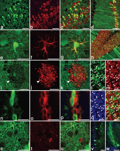
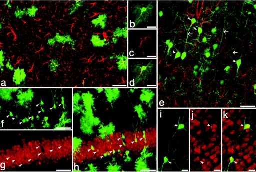
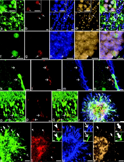
Presently, in vivo methods to efficiently and broadly transduce all major cell types throughout both the central (CNS) and peripheral adult nervous system (PNS) are lacking. In this study, we hypothesized that during early fetal development neural cell populations, including neural stem cells (NSCs), may be accessible for gene transfer via the open neural groove. To test this hypothesis, we injected lentiviral vectors encoding a green fluorescent protein (GFP) marker gene into the murine amniotic cavity at embryonic day 8. This method (i) efficiently and stably transduced the entire nervous system for at least 80% of the lifespan of the mice, (ii) transduced all major neural cell types, and (iii) transduced adult NSCs of the subventricular zone (SVZ) and subgranular zones (SGZs). This simple approach has broad applications for the study of gene function in nervous system development and adult NSCs and may have future clinical applications for treatment of genetic disorders of the nervous system.


Figures

Figure 1
Figure 1
Broad and efficient transduction of the nervous system with a lentiviral construct expressing CMV-GFP (green in all). Expression after E8 intra-amniotic injection is demonstrated in sagittal sections of E12 (a, b) mouse hindbrain, (e) cortex, and (c, d, f–h) dorsal root ganglia, which are located adjacent to myosin (MF20)-positive somites (red, c). Both the (*, d) developing neural tube and (#, d) adjacent dorsal root ganglion are well transduced. CMV-GFP-expressing neurons within the dorsal root ganglion are identified by co-expression of TrkB (red, g, h). Two years after E8 intra-amniotic delivery, strong and stable CMV-GFP expression is present throughout the CNS: macroscopic view of (i) whole brain for CMV-GFP fluorescence, (j) olfactory bulb, (k) hippocampus (NeuN in red), (l) cerebellum, (m) cerebral cortex with a small portion of internal granular layer in lower right corner, (n) spinal cord with strong GFP expression in both anterior and posterior horns as well as frequent GFP+ axons (inset, corticospinal tract) traveling in both motor and sensory tracts, (o) trigeminal nerve innervating the massetter muscle (red), (p) Auerbach's (myenteric) plexus on the small intestine, (q) dorsal root ganglia with spinal nerve, and (r) gross and (s) histological image of transduced adrenal medulla. Red in o and r is background autofluorescence imaged with rhodamine filter set. Within the adrenal, GFP+ cells are found primarily in the medulla and are surrounded by highly autofluorescent adrenal cells (yellow, r) in the zona reticularis. Anti-GFP staining is brown in s. Bar = 20 µm: d, inset of n, o; 100 µm: a–c, e–n, q. CA3, CA3 region of hippocampus; CMV, cytomegalovirus; DG, dentate gyrus; EGL, external granular layer, EPL, external pyramidal layer; GC, granule cells; GFP, green fluorescent protein; GL, glomerular layer; GrL, granular layer; MiL, mitral layer; MoL, molecular layer, PL, Purkinje layer.
Figure 2
Figure 2
Strong and persistent expression of CMV-GFP (green in all panels) in neurons and astrocytes but not microglial cells occurred after E8 intra-amniotic lentiviral delivery. Neurons in the cerebral cortex (red, a–c) and hippocampus (d) that co-express NeuN (red, b–d) are strongly transduced with GFP. Even 2 years after injection, astrocytes (e–g) throughout the CNS stably expressed CMV-GFP as well as the astrocyte-specific protein GFAP (red, f–g). Although rare, an oligodendrocyte (i–k) expressing O4 (red, j,k) in the caudate nucleus appears to express CMV-GFP (i,k) as indicated by the GFP-containing cell body that was centrally located within the oligodendrocyte by three-dimensional confocal analysis. The arrow indicates colocalization of green cytoplasm and O4 staining (arrow) but it is not clear whether the GFP is from a myelinated axon or the oligodendrocyte extension. In intact tissue sections, GFP distributes poorly to oligodendrocyte extensions and O4 often does not stain the cell bodies of oligodendrocytes. Ependymal cells (n–p) expressing CD24 (red, o,p) of the lateral ventricle were also transduced with GFP. However, no GFP-expressing microglial cells (Iba1 staining, red, t,u) were observed. Even at 2 years of age, GFP expression remains strong throughout the CNS as demonstrated by GFP-expressing cerebellar neurons (h; red is NeuN), granular neurons in the cerebellum (l,m,q,r; NeuN is red, m,r; Hoescht-stained nuclei in blue, q), and morphologically distinct Purkinje neurons (v,w; Hoescht-stained nuclei in blue). Bar = 20 µm: e–g, i–w, 50 µm: a–c,v,w; 100 µm: d,h). CMV, cytomegalovirus; CNS, central nervous system; GFP, green fluorescent protein.
Figure 3
Figure 3
GFAP-promoter region driven GFP reveals efficient transduction of astrocytes and neurons. (ak) GFAP-GFP green in all. GFP-expressing astrocytes colocalize with antibody staining for GFAP (red, a–e) in the cortex (a) and hippocampus (b–d). A subset of GFAP-GFP transduced neurons (arrowheads, e–k) expressing GFP in the adult (7 months old) hippocampus (f–h) and caudate (i–k) are identifiable by morphology including long axonal extensions (arrows) and antibody staining for NeuN (red; g,h,j,k). Despite the expression of GFP from GFAP regulatory elements, these neurons lack expression of native GFAP (red, e). Bar = 20 µm: b–d, i–k; 50 µm: a,e–h. GFAP, GFP-expressing astrocyte; GFP, green fluorescent protein.
Figure 4
Figure 4
Evidence for transduction of NSCs after CMV-GFP intra-amniotic injection. GFP expression (green in all) was observed in (a–j, arrows) newly formed neurons that had (red; b,g) divided in adulthood as demonstrated by incorporation of BrdU. Also shown in a–j is the neuronal marker NeuN (tan; d,i,), Hoescht-stained nuclei (blue; c,h) and four-color composite images (e, j). GFP-expressing, new neurons were observed throughout the CNS but were most frequent in the (a–e) olfactory bulbs and (f–j) hippocampi of adult mice. (k–q) GFP-transduced (green), putative NSCs were observed. A single cell expressing GFAP (red; l) that was located immediately underneath ependymal cells expressing CD24 (blue, m) in the subventricular zone expressed a low-level of GFP (lower arrow, k–n). Despite the cytoplasmic localization of GFP (which is occasionally observed in cells weakly expressing GFP), both a hue-based analysis (not shown) and the GFAP+ but GFP cell (upper arrow) provide reassurance of the authenticity of the GFP-signal. Two cells with neuronal morphology (o–q, arrows) co-expressing GFP (o,q) and Nestin (red; p,q), a marker of neural stem and progenitor cells, were observed in the CA3 region of adult hippocampus. Low-density neurosphere cultures (r–v) demonstrate that a single, GFP-expressing cell gives rise to cells of the three major neural lineages: myelin basic protein–expressing oligodendrocytes (red, arrowheads, r,t), GFAP-expressing astrocytes (blue, thin arrows, r,u), and NeuN-expressing neurons (tan, thick arrows, r,v). Representative cells from each image are shown at higher magnification (insets). Although GFP expression appears heterogeneous, all cells within the neurosphere expressed GFP. The limited dynamic range of the detectors in the confocal microscope amplifies the contrast differences between cells, which was likely due to both varying densities of cells in the expanding neurosphere as well as the sensitivity of the CMV promoter to the surrounding chromatin state which varies with cell type. Bar = 10 µm: a–q and insets in t–v; 50 µm: r–v. CA3, CA3 region of hippocampus; CMV, cytomegalovirus; CNS, central nervous system; GFAP, GFP-expressing astrocyte; GFP, green fluorescent protein; NSC, neuronal stem cell.

Similar articles

Cited by

References

    1. Abrous DN, Koehl M., and, Le Moal M. Adult neurogenesis: from precursors to network and physiology. Physiol Rev. 2005;85:523–569. - PubMed
    1. Alvarez-Buylla A, García-Verdugo JM., and, Tramontin AD. A unified hypothesis on the lineage of neural stem cells. Nat Rev Neurosci. 2001;2:287–293. - PubMed
    1. Gage FH. Mammalian neural stem cells. Science. 2000;287:1433–1438. - PubMed
    1. Consiglio A, Gritti A, Dolcetta D, Follenzi A, Bordignon C, Gage FH, et al. Robust in vivo gene transfer into adult mammalian neural stem cells by lentiviral vectors. Proc Natl Acad Sci USA. 2004;101:14835–14840. - PMC - PubMed
    1. Gaiano N, Kohtz JD, Turnbull DH., and, Fishell G. A method for rapid gain-of-function studies in the mouse embryonic nervous system. Nat Neurosci. 1999;2:812–819. - PubMed

Publication types

Substances

LinkOut - more resources