. 2010 Sep;18(9):1667-74.
doi: 10.1038/mt.2010.126. Epub 2010 Jun 22.

Silencing of reporter gene expression in skin using siRNAs and expression of plasmid DNA delivered by a soluble protrusion array device (PAD)

Affiliations

Silencing of reporter gene expression in skin using siRNAs and expression of plasmid DNA delivered by a soluble protrusion array device (PAD)

Emilio Gonzalez-Gonzalez et al. Mol Ther. 2010 Sep.

Abstract

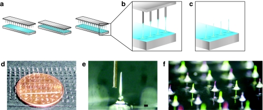
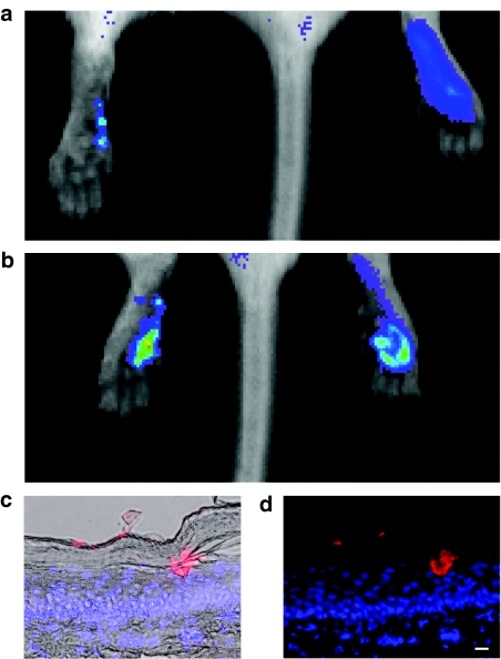
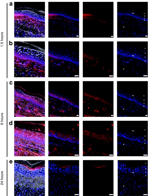
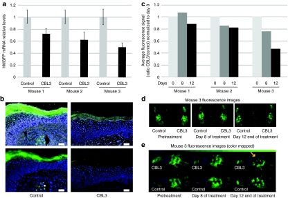
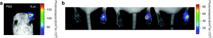
Despite rapid progress in the development of potent and selective small interfering RNA (siRNA) agents for skin disorders, translation to the clinic has been hampered by the lack of effective, patient-friendly delivery technologies. The stratum corneum poses a formidable barrier to efficient delivery of large and/or charged macromolecules including siRNAs. Intradermal siRNA injection results in effective knockdown of targeted gene expression but is painful and the effects are localized to the injection site. The use of microneedle arrays represents a less painful delivery method and may have utility for the delivery of nucleic acids, including siRNAs. For this purpose, we developed a loadable, dissolvable protrusion array device (PAD) that allows skin barrier penetration. The PAD tips dissolve upon insertion, forming a gel-like plug that releases functional cargo. PAD-mediated delivery of siRNA (modified for enhanced stability and cellular uptake) resulted in effective silencing of reporter gene expression in a transgenic reporter mouse model. PAD delivery of luciferase reporter plasmids resulted in expression in cells of the ear, back, and footpad skin as assayed by intravital bioluminescence imaging. These results support the use of PADs for delivery of functional nucleic acids to cells in the skin with an efficiency that may support clinical translation.


Figures

Figure 1
Figure 1
Schematic of fabrication of protrusion array device (PAD) and example images. (a) Pin template and glass slide covered with a thin film of 20% polyvinyl alcohol (PVA) solution (left panel). The pin template is placed in contact with the PVA solution (middle panel). Microneedles are produced by withdrawing the pins as the film is drying, forming fiber-like structures (right panel). (b) Enlarged view of fibers. (c) Protrusions are subsequently trimmed to the desired length and tip shape. (d) PAD supported by a glass substrate, with a penny to show scale. (e) Micrograph of one microneedle after trimming to 1 mm length, showing beveled structure that facilitates skin penetration, with a human hair to show scale (bar = 80 µm). (f) PAD needles loaded alternately with fluorescein (green) and R-phycoerythrin (red).
Figure 2
Figure 2
Imaging of individual microneedle penetration sites in vivo and microneedle plug visualization in skin sections. (a) PADs were loaded with siGLO Red (a fluorescently tagged siRNA mimic, ~20 ng/microneedle) and applied to the left footpad. As a control, 0.5 µg of siGLO Red (in 50 µl PBS) was injected intradermally into the right footpad. Mice were immediately intravitally imaged for fluorescence using the Xenogen IVIS 200 system. Localized fluorescence corresponding to individual microneedle penetration sites was observed following PAD application. (b) The application of four (4 × 1) arrays to the left foot resulted in detection of red fluorescent signal of similar magnitude to the right foot. (c,d) Fluorescence microscopy of 10 µm frozen skin sections showing a microneedle depot loaded with siGLO Red [longer exposure time (data not shown) shows initial release of siGLO Red] demonstrating drug release to the epidermis. Sections were stained with DAPI (blue) to visualize nuclei (bar = 10 µm). (c) Overlay with brightfield image. (d) No brightfield overlay. DAPI, 4,6-diamidino-2-phenylindole; PAD, protrusion array device; PBS, phosphate-buffered saline; siRNA, small interfering RNA.
Figure 3
Figure 3
Fluorescence microscopic analysis of Accell Red siRNA distribution in mouse footpad skin. Transgenic CBL/hMGFP mouse footpads were treated with two (3 × 5) PADs loaded with Accell Red (DY-547-labeled) nontargeting siRNA. Mice were sacrificed at (a,b) 1.5, (c,d) 6, and (e) 24 hours after PAD application and footpad skin sections removed for analysis. Accell Red siRNA (red fluorescence, middle panels) distributes through dermis (d) and epidermis (ep). With this needle design and length, the red fluorescence signal is detected in the basal (b) and spinosum (s) layers at (a,b) 1.5 hours and reaches the granular layer (g) and stratum corneum (sc) at (c,d) 6 hours. Sections were stained with DAPI to visualize nuclei (right panel). Brightfield fluorescence overlay (left panel). Bar = 20 µm. CBL, click beetle luciferase; DAPI, 4,6-diamidino-2-phenylindole; hMGFP, humanized Montastrea green fluorescent protein; PAD, protrusion array device; siRNA, small interfering RNA.
Figure 4
Figure 4
CBL3 Accell siRNA-loaded PADs inhibit hMGFP expression in mouse footpad skin. Transgenic CBL/hMGFP mouse footpads were treated every 2 days with three (3 × 5) PADs loaded with either CBL3 or nonspecific control Accell siRNA for 12 days. (a) RT-qPCR analysis of Tg CBL/hMGFP mice treated with CBL3 siRNA. Total RNA, isolated from paw palm skin of three mice treated with CBL3 siRNA (right footpad) or a nonspecific control siRNA (left footpad), was reverse transcribed and hMGFP mRNA levels quantified by qPCR. The hMGFP levels were normalized to K14 levels (endogenous control). Each bar corresponds to the mean of three replicates. Bars indicate SE. (b) Fluorescence microscopy of frozen skin sections prepared from treated mice. Mice were sacrificed and frozen skin sections (10 µm) prepared. hMGFP expression (or lack thereof) was visualized by fluorescence microscopy of samples from mouse footpads treated with nonspecific (left panel) or CBL3 (right panel) Accell siRNA. Upper panel shows brightfield overlay and bottom panel shows fluorescence only. Bar = 50 µm. Nuclei are visualized by DAPI stain (blue). (c) In vivo quantification of hMGFP fluorescence. Three mice were treated with CBL3 (right paw) or nonspecific (left paw) Accell siRNA and imaged with the CRi Maestro imaging system during treatment. Quantification of the region of interest adjusted to the palm of each mouse was performed using Maestro quantification software after background subtraction. The ratio in the average signal (counts/second/mm2) of CBL3-treated palm and nonspecific-treated palm normalized to day 0 is reported in the graph. (d) hMGFP expression in mouse three imaged during treatment with the CRi Maestro imaging system. The right paw was treated with CBL3 Accell siRNA whereas the left paw was treated with nonspecific control. Images were taken using autoexpose settings and unmixed using previously defined spectra and autofluorescence. The images are pseudocolored green. (e) Enlarged view of mouse three treated paws, color-mapped to a binned black–blue–green color scheme to facilitate comparison of low-intensity regions (see Materials and Methods section). Arrow shows area of signal reduction. Color bar at top shows lookup table assignments corresponding to pixel bit values in increments of 10. CBL, click beetle luciferase; DAPI, 4,6-diamidino-2-phenylindole; hMGFP, humanized Montastrea green fluorescent protein; PAD, protrusion array device; RT-qPCR, reverse transcription quantitative PCR; SE, standard error; siRNA, small interfering RNA; Tg, transgenic.
Figure 5
Figure 5
Analysis of fLuc reporter gene expression following PAD-mediated delivery of expression plasmids to mouse skin. (a) Ear delivery. The ear on the right was treated with a PAD loaded with ~12 ng/microneedle of pGL3-CMV-Luc plasmid (12 microneedles). The ear on the left was treated with the delivery device loaded with PBS vehicle alone. PADs were inserted into the ear for 20 minutes. After 24 hours, luciferase expression was determined following IP luciferin injection by whole animal imaging using the Xenogen IVIS 200 in vivo system (red is the highest expression level, blue lowest). (b) Footpad delivery. Right footpads were treated with PADs (12 microneedles) loaded with luciferase expression plasmid for two consecutive days and analyzed as described above. Left footpads were treated with PADs loaded with PBS vehicle alone (control). fLuc, firefly luciferase; IP, intraperitoneal; PAD, protrusion array device; PBS, phosphate-buffered saline.

References

    1. Uitto J., and, Pulkkinen L. The genodermatoses: candidate diseases for gene therapy. Hum Gene Ther. 2000;11:2267–2275. - PubMed
    1. Smith, FJD, Kaspar, RL, Schwartz, ME, McLean, WHI., and, Leachman, SA.2006. Pachyonychia congenita. GeneReviews < www.genetests.org/profiles/pc >.
    1. Leachman SA, Kaspar RL, Fleckman P, Florell SR, Smith FJ, McLean WH, et al. Clinical and pathological features of pachyonychia congenita. J Investig Dermatol Symp Proc. 2005;10:3–17. - PubMed
    1. Leachman SA, Hickerson RP, Hull PR, Smith FJ, Milstone LM, Lane EB, et al. Therapeutic siRNAs for dominant genetic skin disorders including pachyonychia congenita. J Dermatol Sci. 2008;51:151–157. - PMC - PubMed
    1. Hickerson RP, Smith FJ, Reeves RE, Contag CH, Leake D, Leachman SA, et al. Single-nucleotide-specific siRNA targeting in a dominant-negative skin model. J Invest Dermatol. 2008;128:594–605. - PubMed

Publication types

LinkOut - more resources