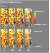
Functional MRI mapping neuronal inhibition and excitation at columnar level in human visual cortex
- PMID: 20571785
- PMCID: PMC2937580
- DOI: 10.1007/s00221-010-2318-z
Functional MRI mapping neuronal inhibition and excitation at columnar level in human visual cortex
Abstract
The capability of non-invasively mapping neuronal excitation and inhibition at the columnar level in human is vital in revealing fundamental mechanisms of brain functions. Here, we show that it is feasible to simultaneously map inhibited and excited ocular dominance columns (ODCs) in human primary visual cortex by combining high-resolution fMRI with the mechanism of binocular inhibitory interaction induced by paired monocular stimuli separated by a desired time delay. This method is based on spatial differentiation of fMRI signal responses between inhibited and excited ODCs that can be controlled by paired monocular stimuli. The feasibility and reproducibility for mapping both inhibited and excited ODCs have been examined. The results conclude that fMRI is capable of non-invasively mapping both excitatory and inhibitory neuronal processing at the columnar level in the human brain. This capability should be essential in studying the neural circuitry and brain function at the level of elementary cortical processing unit.
Figures


Similar articles
-
Mapping iso-orientation columns by contrast agent-enhanced functional magnetic resonance imaging: reproducibility, specificity, and evaluation by optical imaging of intrinsic signal.J Neurosci. 2006 Nov 15;26(46):11821-32. doi: 10.1523/JNEUROSCI.3098-06.2006. J Neurosci. 2006. PMID: 17108155 Free PMC article.
-
Brief visual stimulation allows mapping of ocular dominance in visual cortex using fMRI.Hum Brain Mapp. 2001 Dec;14(4):210-7. doi: 10.1002/hbm.1053. Hum Brain Mapp. 2001. PMID: 11668652 Free PMC article.
-
Ultra-high field fMRI reveals origins of feedforward and feedback activity within laminae of human ocular dominance columns.Neuroimage. 2021 Mar;228:117683. doi: 10.1016/j.neuroimage.2020.117683. Epub 2020 Dec 30. Neuroimage. 2021. PMID: 33385565
-
Development of ocular dominance columns across rodents and other species: revisiting the concept of critical period plasticity.Front Neural Circuits. 2024 Jul 5;18:1402700. doi: 10.3389/fncir.2024.1402700. eCollection 2024. Front Neural Circuits. 2024. PMID: 39036421 Free PMC article. Review.
-
Ocular dominance columns: enigmas and challenges.Neuroscientist. 2009 Feb;15(1):62-77. doi: 10.1177/1073858408327806. Neuroscientist. 2009. PMID: 19218231 Free PMC article. Review.
Cited by
-
Methods for fine scale functional imaging of tactile motion in human and nonhuman primates.Open Neuroimag J. 2011;5:160-71. doi: 10.2174/1874440001105010160. Epub 2011 Nov 18. Open Neuroimag J. 2011. PMID: 22253658 Free PMC article.
-
Denoise Functional Magnetic Resonance Imaging With Random Matrix Theory Based Principal Component Analysis.IEEE Trans Biomed Eng. 2022 Nov;69(11):3377-3388. doi: 10.1109/TBME.2022.3168592. Epub 2022 Oct 19. IEEE Trans Biomed Eng. 2022. PMID: 35439125 Free PMC article.
-
Going Low in a World Going High: The Physiologic Use of Lower Frequency Electron Paramagnetic Resonance.Appl Magn Reson. 2020 Oct;51(9-10):887-907. doi: 10.1007/s00723-020-01261-7. Epub 2020 Oct 18. Appl Magn Reson. 2020. PMID: 33776216 Free PMC article.
-
Cross-species neuroscience: closing the explanatory gap.Philos Trans R Soc Lond B Biol Sci. 2021 Jan 4;376(1815):20190633. doi: 10.1098/rstb.2019.0633. Epub 2020 Nov 16. Philos Trans R Soc Lond B Biol Sci. 2021. PMID: 33190601 Free PMC article.
-
Measurement of ultra-fast signal progression related to face processing by 7T fMRI.Hum Brain Mapp. 2020 May;41(7):1754-1764. doi: 10.1002/hbm.24907. Epub 2020 Jan 10. Hum Brain Mapp. 2020. PMID: 31925902 Free PMC article.
References
-
- Adriany G, Pfeuffer J, Yacoub E, Van de Moortele PF, Shmuel A, Anderson P, Hu X, Vaughan JT, Ugurbil K. A half-volume transmit/receive coil combination for 7 Tesla applications. 9th International society for magnetic resonance in medicine annual meeting; Glasgow, UK. 2001. p. 1097.
-
- Bandettini PA, Wong EC, Hinks RS, Tikofsky RS, Hyde JS. Time course EPI of human brain function during task activation. Magn Reson Med. 1992;25:390–397. - PubMed
-
- Bandettini PA, Jesmanowicz A, Wong EC, Hyde JS. Processing strategies for time-course data sets in functional MRI of the human brain. Magn Reson Med. 1993;30:161–173. - PubMed
-
- Buchert M, Greenlee MW, Rutschmann RM, Kraemer FM, Luo F, Hennig J. Functional magnetic resonance imaging evidence for binocular interactions in human visual cortex. Exp Brain Res. 2002;145:334–339. - PubMed
Publication types
MeSH terms
Grants and funding
- P41 RR08079/RR/NCRR NIH HHS/United States
- P41 RR008079/RR/NCRR NIH HHS/United States
- R01 EB000331/EB/NIBIB NIH HHS/United States
- R01 MH070800/MH/NIMH NIH HHS/United States
- MH70800-01/MH/NIMH NIH HHS/United States
- P30NS057091/NS/NINDS NIH HHS/United States
- R01 NS041262/NS/NINDS NIH HHS/United States
- R56 MH070800/MH/NIMH NIH HHS/United States
- R56 EB000331/EB/NIBIB NIH HHS/United States
- EB00329/EB/NIBIB NIH HHS/United States
- R01 EB000329/EB/NIBIB NIH HHS/United States
- NS41262/NS/NINDS NIH HHS/United States
- EB00331/EB/NIBIB NIH HHS/United States
- P30 NS057091/NS/NINDS NIH HHS/United States
LinkOut - more resources
Full Text Sources
Medical

