. 2010 Aug;28(8):1424-34.
doi: 10.1002/stem.464.

Sin3a is required by sertoli cells to establish a niche for undifferentiated spermatogonia, germ cell tumors, and spermatid elongation

Affiliations

Sin3a is required by sertoli cells to establish a niche for undifferentiated spermatogonia, germ cell tumors, and spermatid elongation

Christopher J Payne et al. Stem Cells. 2010 Aug.

Abstract

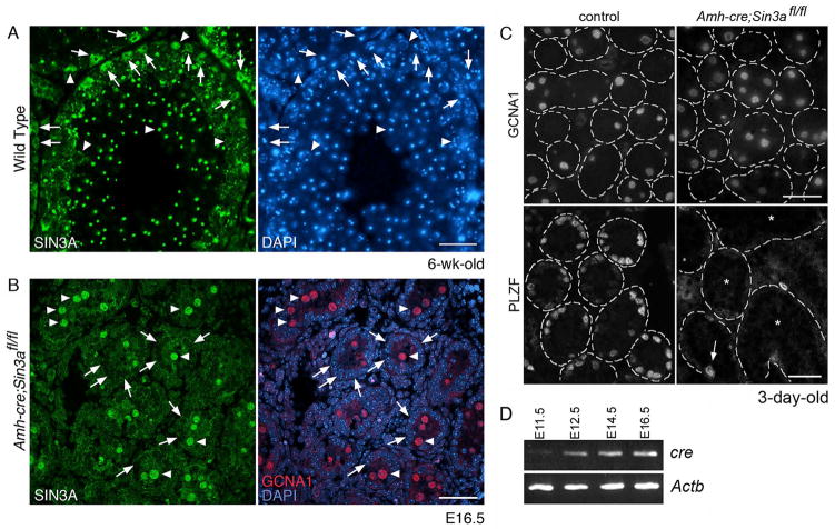
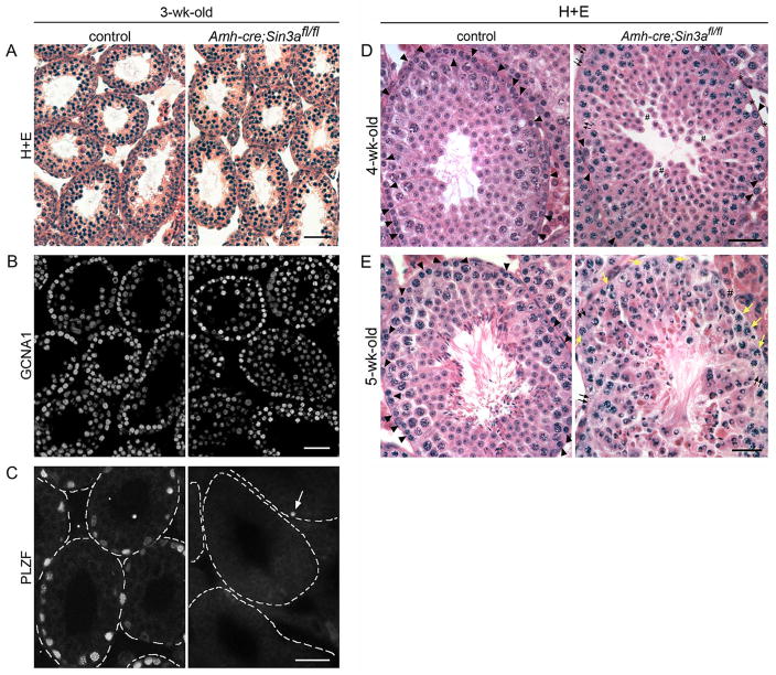
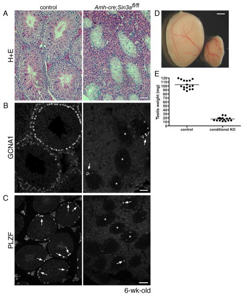
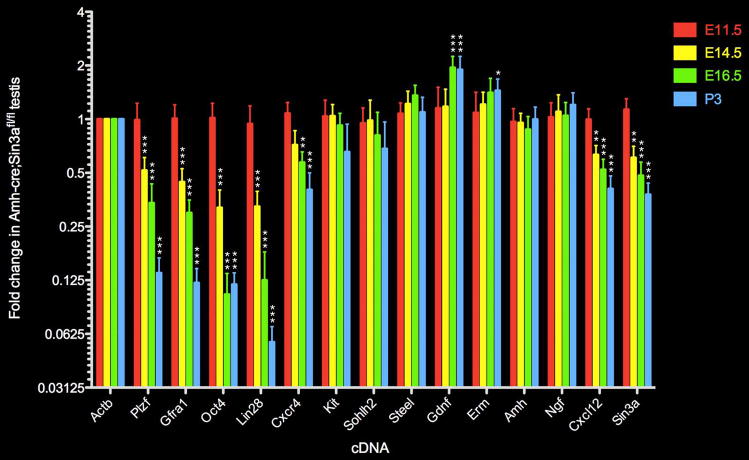
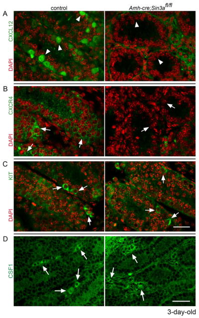
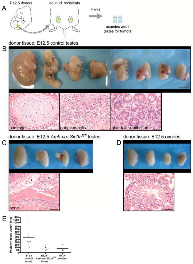
Microenvironments support the maintenance of stem cells and the growth of tumors through largely unknown mechanisms. While cell-autonomous chromatin modifications have emerged as important determinants for self-renewal and differentiation of stem cells, a role for non-cell autonomous epigenetic contributions is not well established. Here, we genetically ablated the chromatin modifier Swi-independent 3a (Sin3a) in fetal Sertoli cells, which partly comprise the niche for male germline stem cells, and investigated its impact on spermatogenic cell fate and teratoma formation in vivo. Sertoli cell-specific Sin3a deletion resulted in the formation of few undifferentiated spermatogonia after birth while initially maintaining spermatogenic differentiation. Stem cell-associated markers Plzf, Gfra1, and Oct4 were downregulated in the mutant fetal gonad, while Sertoli cell markers Steel and Gdnf, which support germ cells, were not diminished. Following birth, markers of differentiating spermatogonia, Kit and Sohlh2, exhibited normal levels, but chemokine-signaling molecules chemokine (C-X-C motif) ligand 12 (CXCL12)/stromal cell-derived factor 1 (SDF1) and chemokine (C-X-C motif) receptor 4 (CXCR4), expressed in Sertoli cells and germ cells, respectively, were not detected. In the juvenile, mutant testes exhibited a progressive loss of differentiating spermatogonia and a block in spermatid elongation, followed by extensive germ cell degeneration. Sertoli cell-specific Sin3a deletion also suppressed teratoma formation by fetal germ cells in an in vivo transplantation assay. We conclude that the epigenome of Sertoli cells influences the establishment of a niche for germline stem cells as well as for tumor initiating cells.


Conflict of interest statement

DISCLOSURE OF POTENTIAL CONFLICTS OF INTEREST

The authors indicate no potential conflicts of interest.

Figures

Figure 1
Figure 1
Sertoli cell-specific Sin3a-deleted testes maintain equivalent numbers of differentiating germ cells after birth, but contain few undifferentiated spermatogonia. (A): Cross-section of a 6-wk-old wild type testis immunostained with anti-SIN3A antibody (left) and labeled with DAPI (right). Arrows identify Sertoli cell nuclei; arrowheads denote germ cell nuclei. (B): Cross-section of an E16.5 Amh-cre;Sin3afl/fl testis co-immunostained with anti-SIN3A antibody (left) and anti-germ cell nuclear antigen (GCNA; right, red), and labeled with DAPI (right, blue). Arrows identify Sertoli cell nuclei; arrowheads denote gonocyte nuclei. (C): Cross-sections of 3-day-old control Amh-cre;Sin3a+/+ (left) and mutant Amh-cre;Sin3afl/fl (right) testes immunostained with anti-GCNA1 antibody (top) and anti-PLZF antibody (bottom). The outlines of distinct seminiferous tubules are demarcated with dashed lines (representing basement membranes). Note the appearance of equivalent numbers of GCNA1-positive germ cells. Arrow (bottom right) identifies a single PLZF-positive spermatogonium in the field of view. Asterisks denote seminiferous tubules completely devoid of undifferentiated spermatogonia. (D): RT-PCR products for cre (Amh-cre) and Actb genes expressed in E11.5 gonads and E12.5, E14.5, and E16.5 testes. Note the increasing levels of cre that correspond with developmental progression. Scale bars represent 50μm (A), 25μm (B), 25 μm (C, top panels), and 50μm (C, bottom panels).
Figure 2
Figure 2
Juvenile Amh-cre;Sin3afl/fl testes exhibit a progressive loss of spermatogonia and a block in spermatid elongation. (A): Cross-sections of 3-wk-old control Amh-cre;Sin3a+/+ (left) and mutant Amh-cre;Sin3afl/fl (right) testes stained with hematoxylin and eosin (H+E). (B): Cross-sections of 3-wk-old control (left) and conditional knockout (right) testes immunostained with anti-GCNA1 antibody. Note the appearance of equivalent numbers of GCNA1-positive germ cells. (C): Cross-sections of 3-wk-old control (left) and conditional knockout (right) testes immunostained with anti-PLZF antibody. The outlines of distinct seminiferous tubules are demarcated with dashed lines (representing basement membranes). Arrow identifies a single PLZF-positive spermatogonium in the field of view. (D): Cross-sections of 4-wk-old control (left) and conditional knockout (right) testes stained with H+E. Arrowheads identify spermatogonia that reside along the basement membrane; small black arrows indicate regions of the basal compartment devoid of germ cells; asterisks denote empty spaces suggestive of where spermatogonia used to reside; pound signs mark the adlumenal compartment where round spermatids exhibit increased cell separation from each other. (E): Cross-sections of 5-wk-old control (left) and conditional knockout (right) testes stained with H+E. Arrowheads identify spermatogonia on the basement membrane; small black arrows indicate regions devoid of germ cells; small yellow arrows denote meiotic spermatocytes that now reside along the basement membrane; pound sign marks round spermatids occupying the basal compartment. Note the absence of elongated spermatids in the Amh-cre;Sin3afl/fl testis, and the abnormal-looking round spermatid nuclei with clear zones, indicative of cell degeneration. Scale bars represent 50μm (A–E).
Figure 3
Figure 3
Adult Amh-cre;Sin3afl/fl testes are depleted of germ cells. (A): Cross-sections of 6-wk-old control Amh-cre;Sin3a+/+ (left) and mutant Amh-cre;Sin3afl/fl (right) testes stained with hematoxylin and eosin. Note the reduced diameters of Amh-cre;Sin3afl/fl seminiferous tubules. (B): Cross-sections of adult control (left) and conditional knockout (right) testes immunostained with anti-GCNA1 antibody. Arrows identify GCNA1-positive germ cells in two seminiferous tubules; asterisks denote three seminiferous tubules completely devoid of germ cells. (C): Cross-sections of adult control (left) and conditional knockout (right) testes immunostained with anti-PLZF antibody. Arrows identify PLZF-positive spermatogonia; asterisks denote three seminiferous tubules in the mutant cross-section completely devoid of undifferentiated spermatogonia. (D): Representative examples of 6-wk-old control Amh-cre;Sin3a+/+ (left) and mutant Amh-cre;Sin3afl/fl (right) testes. (E): Weights of 6-wk-old control (left) and conditional knockout (right) testes. Scale bars represent 50μm (A, B), 75μm (C), and 1mm (D).
Figure 4
Figure 4
Levels of transcripts specific to gonocytes and undifferentiated spermatogonia are diminished following Sin3a deletion in fetal Sertoli cells. Quantitative real time RT-PCR analysis depicting the fold change in expression of selected gonocyte and undifferentiated spermatogonia-specific genes (Plzf, Gfra1, Oct4, Lin28), differentiating germ cell-specific genes (Kit, Sohlh2), Sertoli cell genes (Steel, Gdnf, Amh, Cxcl12, Erm, Ngf), germ cell gene Cxcr4, germline and soma gene Sin3a, and internal control gene Actb in E11.5 (red), E14.5 (yellow), E16.5 (green), and P3 (blue) mutant Amh-cre;Sin3afl/fl testes relative to gene expression levels in control Amh-cre;Sin3a+/+ testes (given an arbitrary value of 1 for all genes in control testes, not shown in this plot). Data are represented as mean values +/− SEM. Statistical comparisons with control testis-expressed genes: *p<0.05; **p<0.01; ***p<0.001.
Figure 5
Figure 5
Chemokine CXCL12 and its receptor CXCR4 are not detected in neonatal Amh-cre;Sin3afl/fl testes, while the KIT receptor and cytokine CSF1 distribute normally. Cross-sections of 3-day-old control Amh-cre;Sin3a+/+ (left) and mutant Amh-cre;Sin3afl/fl (right) testes, immunostained with (A): anti-CXCL12 antibody (green), (B): anti-CXCR4 antibody (green), (C): anti-KIT antibody (green), (D): anti-CSF1 antibody (green) and labeled with DAPI (red, A–C). Arrowheads denote Sertoli cell cytoplasm and extracellular regions; arrows identify spermatogonia (A–C) and clusters of Leydig cells in the interstitium (D). Note the lack of CXCL12 and CXCR4 in the seminiferous tubules of the conditional knockout testes. Scale bars represent 25μm (A-C) and 50μm (D).
Figure 6
Figure 6
Transplanted fetal Amh-cre;Sin3afl/fl testes suppress the formation of adult testicular germ cell tumors. (A): Schematic diagram of the transplantation assay. Gonads from E12.5 embryos (yellow) were isolated and transplanted to the testicular parenchyma of age-matched adults (blue). Four weeks later, adult testes were removed and examined for teratomas. (B, top panel): Adult testes that received E12.5 control testes as donor tissue. Testes are irregularly enlarged, containing multilobular tumors. (B, bottom panels): Tumors from all of the testes are comprised of well-differentiated tissue arising from all three embryonic germ layers (mesoderm, endoderm, neuroectoderm), consistent with teratomas. (C, top panel): Adult testes that received E12.5 Amh-cre;Sin3afl/fl testes as donor tissue, exhibiting modest enlargement. (C, bottom panel): Representative example of a teratoma from one testis containing newly formed bone. Asterisks denote degenerating seminiferous tubules. (D, top panel): Adult testes that received E12.5 ovaries as donor tissue. (D, bottom panel): Seminiferous tubule containing spermatogenic cells. (E): Weights of adult testes that received E12.5 control testes, E12.5 Amh-cre;Sin3afl/fl testes, and E12.5 ovaries as donor tissue. Scale bars represent 5 mm in the top panels (B–D), and 50μm in the bottom panels (B–D).

Similar articles

Cited by

References

    1. Schofield R. The relationship between the spleen colony-forming cell and the haemopoietic stem cell. Blood Cells. 1978;4:7–25. - PubMed
    1. Fuchs E, Segre JA. Stem cells: a new lease on life. Cell. 2000;100:143–155. - PubMed
    1. Mills JC, Gordon JI. The intestinal stem cell niche: there grows the neighborhood. Proc Natl Acad Sci U S A. 2001;98:12334–12336. - PMC - PubMed
    1. Doetsch F. A niche for adult neural stem cells. Curr Opin Genet Dev. 2003;13:543–550. - PubMed
    1. Shinohara T, Orwig KE, Avarbock MR, et al. Remodeling of the postnatal mouse testis is accompanied by dramatic changes in stem cell number and niche accessibility. Proc Natl Acad Sci U S A. 2001;98:6186–6191. - PMC - PubMed

Publication types

Substances