. 2010 Aug;28(8):1355-67.
doi: 10.1002/stem.465.

ATP-sensitive K(+) channel-deficient dilated cardiomyopathy proteome remodeled by embryonic stem cell therapy

Affiliations

ATP-sensitive K(+) channel-deficient dilated cardiomyopathy proteome remodeled by embryonic stem cell therapy

Jelena Zlatkovic-Lindor et al. Stem Cells. 2010 Aug.

Abstract

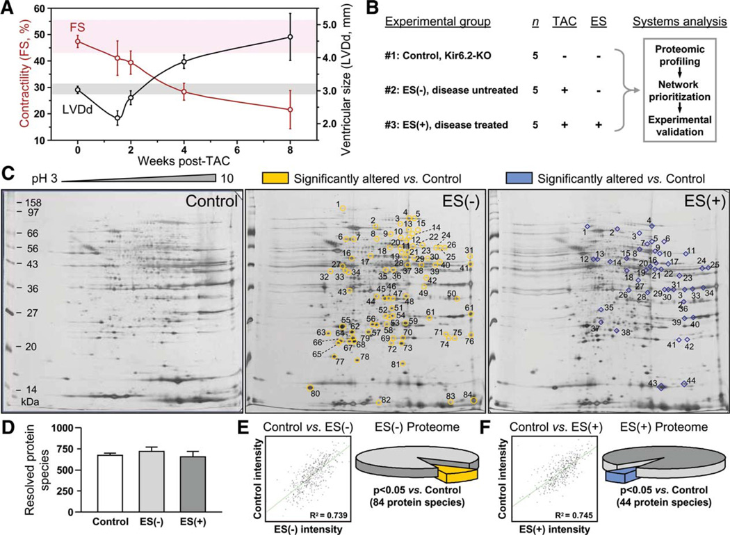
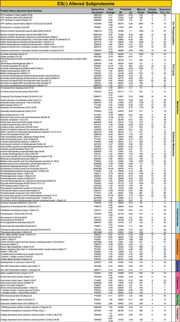
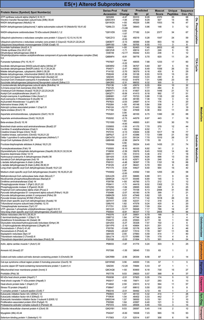
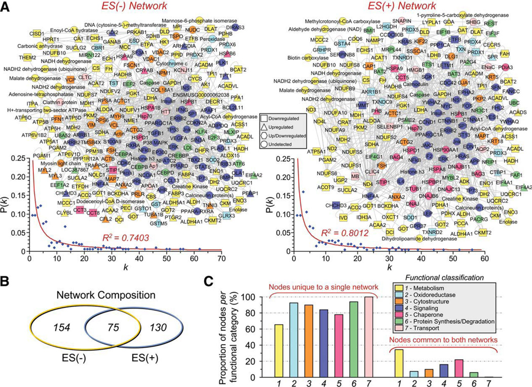
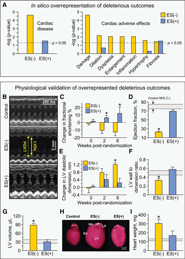
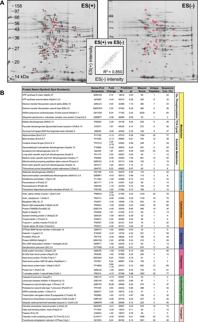
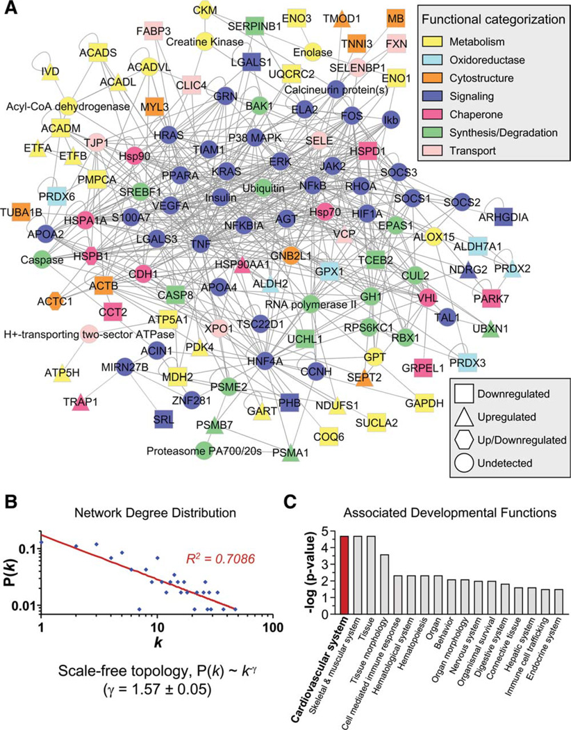
Transplantation of pluripotent stem cells has proven beneficial in heart failure, yet the proteomic landscape underlying repair remains largely uncharacterized. In a genetic model of dilated cardiomyopathy elicited by pressure overload in the KCNJ11 (potassium inwardly rectifying channel, subfamily J, member 11) null mutant, proteome-wide profiles were here resolved by means of a systems approach prior to and following disease manifestation in the absence or presence of embryonic stem cell treatment. Comparative two-dimensional gel electrophoresis revealed a unique cardiomyopathic proteome in the absence of therapy, remodeled in response to stem cell treatment. Specifically, linear ion trap quadrupole-Orbitrap mass spectrometry determined the identities of 93 and 109 differentially expressed proteins from treated and untreated cardiomyopathic hearts, respectively. Mapped protein-protein relationships and corresponding neighborhoods incorporated the stem cell-dependent subproteome into a nonstochastic network with divergent composition from the stem cell-independent counterpart. Stem cell intervention produced a distinct proteome signature across a spectrum of biological processes ranging from energetic metabolism, oxidoreductases, and stress-related chaperones to processes supporting protein synthesis/degradation, signaling, and transport regulation, cell structure and scaffolding. In the absence of treatment, bioinformatic interrogation of the disease-only proteome network prioritized adverse cardiac outcomes, ablated or ameliorated following stem cell transplantation. Functional and structural measurements validated improved myocardial contractile performance, reduced ventricular size and decreased cardiac damage in the treated cohort. Unbiased systems assessment unmasked "cardiovascular development" as a prioritized biological function in stem cell-reconstructed cardiomyopathic hearts. Thus, embryonic stem cell treatment transformed the cardiomyopathic proteome to demote disease-associated adverse effects and sustain a procardiogenic developmental response, supplying a regenerative substrate for heart failure repair.


Conflict of interest statement

Disclosure of Potential Conflicts of Interest.

The authors indicate no potential conflicts of interest.

Figures

Figure 1
Figure 1
Stem cell intervention remodels heart proteome in genetic dilated cardiomyopathy. (A): Malignant dilated cardiomyopathy in KATP channel-deficient (Kir6.2-KO) hearts under stress imposed by TAC characterized by decreased contractility (FS) and increased LVDd following initial compensatory hypertrophy observed at 1.5 weeks post-TAC. Shaded background indicates 95% confidence interval for FS (pink) and LVDd (gray) in age- and sex-matched Kir6.2-KO (Control) hearts. (B): The experimental protocol involved systems analysis combining proteomic comparison of left ventricular (LV) extracts obtained 8 weeks post-TAC from three separate experimental groups, unstressed Kir6.2-KO (#1 - Control), disease untreated TAC Kir6.2-KO [#2 – ES(−)] and disease treated TAC Kir6.2-KO [#3 – ES(+)] treated by 6 weeks of R1 embryonic stem cell therapy, followed by network analysis and in silico prioritization of proteomic findings in conjunction with in vivo and ex vivo functional validation. (C): Representative silver stained two-dimensional gels (pH 3–10 IEF, 12.5% SDS-PAGE) of LV tissue extracts (100 µg protein) from unstressed Kir6.2-KO (control), and from aortic-constricted untreated [ES(−)] or stem cell-treated [ES(+)] counterparts. Spots identified as differentially expressed relative to those in unstressed hearts are circled and numbered on ES(−) and ES(+) gels. (D): Gel reproducibility demonstrated by no significant difference in number of resolved protein species. (E, F): Densitometric spot quantitation, with scatter plots of average normalized intensities of matching protein spots showing correlation for control versus ES(−) and control versus ES(+) gels, and pie charts indicating significant differences in 84 and 44 protein species for control versus ES(−) and control versus ES(+), respectively (p < .05). Abbreviations: ES, embryonic stem cells; FS, fractional shortening; LVDd, left ventricular end-diastolic dimension; TAC, transverse aortic constriction.
Figure 2
Figure 2
Identity of cardiomyopathy-induced subproteome. The 109 significantly altered proteins induced by progressive cardiomyopathy [ES(−) altered subproteome] and identified by LTQ-Orbitrap MS/MS analysis, were functionally categorized and color-coded by Swiss-Prot ontological annotation. Protein names are listed with their symbol (Swiss-Prot gene abbreviation) and spot numbers to locate corresponding 2D gel position(s) in Figure 1. Mascot score, number of unique identified peptides, % sequence cov. (coverage), predicted Mr and pI for each protein (following expected post-translational processing, for example, removal of a mitochondrial signal peptide), and fold change of ES(−) over control are indicated. For proteins detected in more than one spot, maximum score and corresponding number of unique peptides are reported. Fold change was calculated as described in experimental procedures, and for proteins detected in both increasing and decreasing spots (*), both values are indicated. Abbreviations: 2D, two-dimensional; CDGSH, Unique 39 amino acid CDGSH domain [C-x-C-x2-(S/T)-x3-P-x-C-D-G-(S/A/T)-H]; ES, Embryonic stem cells; LTQ, Linear ion trap quadrupole; MS/MS, Tandem mass spectrometry; Ox., oxidative; Syn./Deg., protein synthesis/degradation; TCA cycle, tricarboxylic acid cycle.
Figure 3
Figure 3
Identity of cardiomyopathic subproteome following stem cell therapy. The 93 significantly altered proteins induced by stem cell therapy in the setting of dilated cardiomyopathy [ES(+) altered subproteome], and identified by LTQ-Orbitrap MS/MS analysis, were functionally categorized and color-coded by Swiss-Prot ontological annotation. Protein names are listed with their symbol (Swiss-Prot gene abbreviation) and spot numbers to locate corresponding 2D gel position(s) in Figure 1. Mascot score, number of unique identified peptides, % sequence cov. (coverage), predicted Mr and pI for each protein (following expected post-translational processing, for example, removal of a mitochondrial signal peptide), and fold change of ES(+) over control are indicated. For proteins detected in more than one spot, maximum score and corresponding number of unique peptides are reported. Fold change was calculated as described in experimental procedures, and for proteins detected in both increasing and decreasing spots (*), both values are indicated. Abbreviations: 2D, two-dimensional; Ox., oxidative; Syn./Deg., protein synthesis/ degradation; TCA cycle, tricarboxylic acid cycle.
Figure 4
Figure 4
Stem cell therapy transforms the cardiomyopathy-associated protein interaction network. (A): The 109 ES(−) and 93 ES(+) differentially expressed proteins were submitted to IPA as focus nodes, generating broader interaction networks of 229 and 205 proteins, respectively. Proteins are designated by symbols corresponding to Swiss-Prot gene abbreviations or by family name for nodes representing protein families, and are colored by the functional ontology detailed in Figures 2 and 3, with node shape indicating directionality of focus protein expression change (legend) and nodes common to both networks maintained in the same spatial location. Plots of degree distribution [P(k)] versus degree (k) followed power law distributions, where P(k) ~k−γ, with γ= 1.55 ± 0.04 for ES(−) and 1.57 ± 0.04 for ES(+) networks, respectively, indicating scale-free, nonstochastic network architecture. (B): Limited overlap was found between ES(−) and ES(+) networks (total unique and common nodes indicated), with (C) the majority of proteins from each functional category residing within only one of the two networks. Abbreviations: ES, embryonic stem cells; P(k), degree distribution; k, node degree.
Figure 5
Figure 5
Stem cell-dependent demotion of disease-associated adverse effects. (A): Bioinformatic interrogation of the ES(−) and ES(+) networks within Ingenuity Pathways Knowledge Base for deleterious outcomes indicated that significant overrepresentation of “Cardiac disease” within the ES(−) network was reduced by three orders of magnitude in the ES(+) network (left panel). Screening against Ingenuity toxicological pathways further indicated seven adverse effects in the ES(−) subproteome, all of which were cardiac specific, that is, damage, dilation, dysplasia, enlargement, inflammation, hypertrophy and fibrosis, and nearly all were abolished in the ES(+) subproteome (right panel). (B): Echocardio-graphic measurements indicated that (C) LV fractional shortening, (D) LV ejection fraction, (E) LV diastolic dimension, (F) LV wall to dimension ratio, and (G) LV end-diastolic volume were all significantly worsened by pressure overload but returned to prestress levels following 6 weeks of cell therapy. (H–I): Measurement of hearts on autopsy indicated that the significant increase in heart mass imposed by pressure overload was reversed by cell therapy. Collectively, these parameters demonstrated cell therapy-induced improvement in myocardial contractile performance, reduction in LV size and decreased cardiac damage (*, p < .01 ES(−) versus ES(+) and ES(−) versus control, with control indicated by 95% C.I. in gray as noted in panel (D). Abbreviations: C.I., confidence intervals; ES, embryonic stem cells; LA, left atrium; LV, left ventricle; LVDd, left ventricular end-diastolic dimension; LVDs, left ventricular end-systolic dimension; RA, right atrium; RV, right ventricle.
Figure 6
Figure 6
Stem cell therapy-specific subproteome. (A): Comparison of stem cell-treated [ES(+)] versus untreated [ES(−)] left ventricular tissue extracts by two-dimensional (2D) electrophoresis. Differentially expressed spots isolated for identification by LTQ-Orbitrap mass spectrometric analysis are circled, and numbered on the ES(+) gel. Inset: Gel-to-gel reproducibility indicated by correlation of scatter plot for average normalized densitometric intensities of matching protein spots from ES(+) versus ES(−) gels. (B): Identities of the 61 proteins significantly altered by cell therapy are listed with their symbol (Swiss-Prot gene abbreviation) and spot numbers to locate corresponding 2D gel position(s) in panel (A). Mascot score, number of unique identified peptides, % sequence cov. (coverage), predicted Mr and pI for each protein (following expected post-translational processing, for example, removal of a mitochondrial signal peptide), and fold change in ES(+) versus ES(−) are indicated. For proteins detected in more than one spot, maximum score and corresponding number of unique peptides are reported. Fold change was calculated as described in experimental procedures, and for proteins detected in both increasing and decreasing spots (*), both values are indicated. Abbreviations: ES, embryonic stem cells; Ox., oxidative; TCA cycle, tricarboxylic acid cycle; SAPK, stress activated protein kinase.
Figure 7
Figure 7
Cardiovascular system development prioritized in stem cell therapy-specific network. (A): Submission to Ingenuity Pathways Analysis (IPA) of differentially expressed proteins in the ES(+)-treated hearts generated a 120-protein interaction network, functionally categorized and color-coded by Swiss-Prot ontological annotation (upper legend), with proteins designated by symbols corresponding to Swiss-Prot gene abbreviations or by family name for nodes representing protein families, and shape indicating directionality of focus protein expression change (lower legend). (B): A log-log plot of ES(+)-specific degree distribution [P(k)] versus degree (k) followed a power law distribution, indicating scale-free, nonstochastic network architecture. (C): Bioinformatic interrogation of IPA for associated developmental functions revealed prioritization of cardiovascular system development in the ES(+)-de-pendent proteome in response to cell therapy. Abbreviations: P(k), degree distribution; k, node degree.

Similar articles

Cited by

References

    1. Towbin JA, Bowles NE. The failing heart. Nature. 2002;415:227–233. - PubMed
    1. Hershberger RE, Lindenfeld J, Mestroni L, et al. Genetic evaluation of cardiomyopathy: A Heart Failure Society of America practice guideline. J Card Fail. 2009;15:83–97. - PubMed
    1. Luk A, Ahn E, Soor GS, et al. Dilated cardiomyopathy: A review. J Clin Pathol. 2009;62:219–225. - PubMed
    1. Chien KR. Genomic circuits and the integrative biology of cardiac diseases. Nature. 2000;407:227–232. - PubMed
    1. Ahmad F, Seidman JG, Seidman CE. The genetic basis for cardiac remodeling. Annu Rev Genomics Hum Genet. 2005;6:185–216. - PubMed

Publication types