. 2010 Jul;64(1):47-53.
doi: 10.1002/mrm.22376.

Multishot PROPELLER for high-field preclinical MRI

Affiliations

Multishot PROPELLER for high-field preclinical MRI

Prachi Pandit et al. Magn Reson Med. 2010 Jul.

Abstract

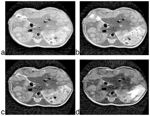
With the development of numerous mouse models of cancer, there is a tremendous need for an appropriate imaging technique to study the disease evolution. High-field T(2)-weighted imaging using PROPELLER (Periodically Rotated Overlapping ParallEL Lines with Enhanced Reconstruction) MRI meets this need. The two-shot PROPELLER technique presented here provides (a) high spatial resolution, (b) high contrast resolution, and (c) rapid and noninvasive imaging, which enables high-throughput, longitudinal studies in free-breathing mice. Unique data collection and reconstruction makes this method robust against motion artifacts. The two-shot modification introduced here retains more high-frequency information and provides higher signal-to-noise ratio than conventional single-shot PROPELLER, making this sequence feasible at high fields, where signal loss is rapid. Results are shown in a liver metastases model to demonstrate the utility of this technique in one of the more challenging regions of the mouse, which is the abdomen.


Figures

Figure 1
Figure 1
k-space trajectory (a) traversed by PROPELLER and the pulse sequence diagram (b) for the same as implemented in the EPIC Lx12M4 platform.
Figure 2
Figure 2
Echo number to blade position correspondence in (a) conventional PROPELLER and 2-shot PROPELLER with (b) early, (c) intermediate, and (d) late acquisition order. The echo number increases as the echo amplitude decreases. In the 2-shot case, the bold and the dotted lined represent the two echo trains that make up a single blade. Effective echo time is defined as the time at which the echo at the k-space center is acquired.
Figure 3
Figure 3
Reconstruction flow for 2-shot PROPELLER in small animals.
Figure 4
Figure 4
Comparison between standard Cartesian fast spin echo (a, b), conventional PROPELLER (c, d), and 2-shot PROPELLER (e, f). Multi-slice datasets with similar imaging parameters were acquired with all three sequences (TR/TE = 3s/70ms, imaging time = 30 minutes). This figure shows two identical slices, one through the liver (a, c, e) and one through the kidney (b, d, f). The arrow points to ghosting artifact in the vena cava seen in single-shot PROPELLER, but absent in the 2-shot scheme.
Figure 5
Figure 5
T2-weighted images of kidneys in a free-breathing mouse. Effective echo times were varied by changing the ETL of the scans (a) TE = 34ms, ETL = 8, (b) TE = 48ms, ETL = 12, (c) TE = 62ms, ETL = 16, (d) CNR between the outer renal medulla (region 1) and the renal cortex (region 2) and inner medulla (region 3).
Figure 6
Figure 6
Liver tumor images from a free-breathing mouse acquired over a range of increasing echo times. 117μm in-plane, 1mm slice thickness (21 slices), TR = 2.5s, BW = 125 KHz, ESP = 6.856ms, imaging time ~ 40 minutes. (a) TE = 27ms, ETL = 6, (b) TE = 41ms, ETL = 10, (c) TE = 55ms, ETL = 14, (d) TE = 69ms, ETL = 18.

References

    1. Marzola P, Osculati F, Sbarbati A. High field MRI in preclinical research. Eur J Radiol. 2003;48(2):165–170. - PubMed
    1. Hedlund LW, Cofer GP, Owen SJ, Allan Johnson G. MR-compatible ventilator for small animals: computer-controlled ventilation for proton and noble gas imaging. Magn Reson Imaging. 2000;18(6):753–759. - PubMed
    1. Pipe JG. Motion correction with PROPELLER MRI: application to head motion and free-breathing cardiac imaging. Magn Reson Med. 1999;42(5):963–969. - PubMed
    1. DiFrancesco MW, Rasmussen JM, Yuan W, Pratt R, Dunn S, Dardzinski BJ, Holland SK. Comparison of SNR and CNR for in vivo mouse brain imaging at 3 and 7 T using well matched scanner configurations. Med Phys. 2008;35(9):3972–3978. - PMC - PubMed
    1. Malisch TW, Hedlund LW, Suddarth SA, Johnson GA. MR microscopy at 7. 0 T: effects of brain iron. J Magn Reson Imaging. 1991;1(3):301–305. - PubMed

Publication types