Review
. 2010 May;69(5):427-42.
doi: 10.1111/j.1365-2125.2010.03629.x.

Role of voltage-gated sodium, potassium and calcium channels in the development of cocaine-associated cardiac arrhythmias

Affiliations
Review

Role of voltage-gated sodium, potassium and calcium channels in the development of cocaine-associated cardiac arrhythmias

Michael E O'Leary et al. Br J Clin Pharmacol. 2010 May.

Abstract

Cocaine is a highly active stimulant that alters dopamine metabolism in the central nervous system resulting in a feeling of euphoria that with time can lead to addictive behaviours. Cocaine has numerous deleterious effects in humans including seizures, vasoconstriction, ischaemia, increased heart rate and blood pressure, cardiac arrhythmias and sudden death. The cardiotoxic effects of cocaine are indirectly mediated by an increase in sympathomimetic stimulation to the heart and coronary vasculature and by a direct effect on the ion channels responsible for maintaining the electrical excitability of the heart. The direct and indirect effects of cocaine work in tandem to disrupt the co-ordinated electrical activity of the heart and have been associated with life-threatening cardiac arrhythmias. This review focuses on the direct effects of cocaine on cardiac ion channels, with particular focus on sodium, potassium and calcium channels, and on the contributions of these channels to cocaine-induced arrhythmias. Companion articles in this edition of the journal examine the epidemiology of cocaine use (Wood & Dargan) and the treatment of cocaine-associated arrhythmias (Hoffmann).


Figures
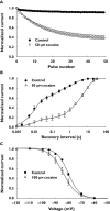
Figure 1
Figure 1
Properties of cocaine inhibition of cardiac Na channels. Effects of cocaine on cardiac (Nav1.5) Na channels heterologously expressed on Xenopus oocytes were investigated. (A) Use-dependent inhibition induced by applying depolarizing pulses at a frequency of 5 Hz. (B) Recovery from inactivation measured in the absence and presence of cocaine. In control experiments the channels recovered with time constants of 11 and 189 ms. After application of cocaine the majority of the channels (77%) recovered with a time constant of 7.5 s. (C) Steady-state inactivation was measured by applying depolarizing prepulses between −120 and −60 mV for 60s. The smooth curves are fits to a Boltzmann function with midpoints of −80 mV for control and −84 mV after application of cocaine. Figure reproduced with permission from O'Leary [47]
Figure 2
Figure 2
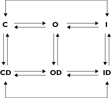
State-dependent drug binding to Na channels. Modulated receptor model describing the state-dependent binding of local anaesthetics. C, O and I represent the closed, open and inactivated states of Na channels, respectively. CD, OD and ID are the drug-modified (D) equivalents. Anaesthetics generally bind with low affinity to closed channels (C) and high affinity to the open (O) and inactivated (I) states. Drugs rapidly access the binding site on open channels (O→OD) through the aqueous pore. Access to the binding site on the closed (C→CD) and inactivated (I→ID) states is through an intrinsically slower hydrophobic pathway
Figure 3
Figure 3
Cocaine block of an inactivation-deficient Na channel mutant. The inactivation-deficient mutant was constructed by replacing hydrophobic residues of the interdomain D3-D4 linker of the cardiac (Nav1.5) Na channel with glutamines (IFM→QQQ) [64]. The mutant channels were heterogously expressed in Xenopus oocytes and Na currents recorded using two-electrode voltage clamp. (A) Currents of non-inactivating mutant before (CTRL) and after application of cocaine (25–150 µm). (B) The decay of the current was fitted with an exponential function and the apparent blocking rate (1/τ) plotted vs. the cocaine concentration. The straight line predicts a KD for cocaine binding at −10 mV of 122 µm. Figure reprinted with permission from O'Leary & Chahine [47]
Figure 4
Figure 4
Schematic of cocaine-induced cardiotoxicity related to Na channel inhibition. Cocaine stabilizes Na channels in inactivated states that do not conduct Na current. Na channel inhibition slows the rapid upstroke of the AP (Phase 0 depolarization), an important determinant of intracardiac conduction. Slowed conduction decreases myocardial contractility leading to depressed left ventricular function and haemodynamic impairment. Prolongation of ventricular depolarization (QRS interval) exposes the myocardium to potentially lethal re-entrant arrhythmias. Combining Na channel inhibition with other pro-arrhythmic electrical disturbances, such as the inhibition of delayed rectifier hERG (long QT intervals) or l-type calcium channels, further increases the likelihood of arrhythmias and sudden cardiac death. Cocaine-induced ischaemia caused by enhanced sympathomimetic stimulation of coronary vasculature can lead to myocardial damage that further potentiates Na channel inhibition and cardiac arrhythmias. Cocaine binding is enhanced at rapid heart rates due to use-dependent inhibition, under conditions where the resting membrane potential is depolarized due to high-affinity binding to inactivated states and during episodes of acidosis, which stabilizes cocaine in its more potent positively charged form
Figure 5
Figure 5
Role of IKr in ventricular action potential repolarization. Schematic diagram showing the profile of IKr (lower trace) during the ventricular action potential (upper trace). Outward current flow via IKr increases throughout the plateau phase of the action potential (marked by filled arrows), peaking before terminal repolarization, during which IKr declines (marked by open arrow)
Figure 6
Figure 6
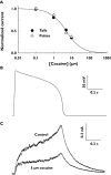
Inhibition of IhERG by cocaine. (A) shows concentration–response relations for inhibition by cocaine of IhERG recorded from hERG-expressing tsA201 cells. IhERG was elicited by depolarization to +20 mV and tail currents were observed on repolarization to −80 mV. Concentration–response relations were similar whether IhERG amplitude was measured for currents during the depolarizing pulse or for current tails. The calculated IC50 value was 5.6 ± 0.4 µm. (B) shows ventricular action potential waveform used for AP clamp experiments in which effects of cocaine on IhERG from ts201 cells were studied. (C) IhERG in standard external solution (control) and following exposure to 5 µm cocaine. Data are reproduced from O'Leary [128] with permission
Figure 7
Figure 7
Schematic diagram showing amino-acid residues on hERG implicated in cocaine binding. The figure shows a vertical cross section through two of the four subunits that comprise functional hERG channels, focusing on the S6 and pore helical regions implicated in drug binding. The selectivity filter of the channel and direction of outward flux (as would occur physiologically through open channels) of K+ ions are also shown. Aromatic residues Y652 and F656 are implicated in the binding of a range of drugs and their mutation influences cocaine binding [133]. T623 is also critical for cocaine binding, whilst mutation of S624 also influences observed potency of inhibition, though to a less marked extent [133]. Mutation of the nearby S620 (not shown) to threonine, but not cysteine has also been shown to influence potency of cocaine inhibition of IhERG[133]
Figure 8
Figure 8
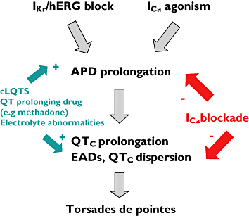
Schematic diagram showing links between hERG, l-type Ca current and cocaine-induced arrhythmia. Vertical ‘information flow’ (downward arrows) shows consequences of IKr/IhERG inhibition, namely prolongation of ventricular action potential duration (APD) and consequent QTc prolongation and QTc dispersion at the intact tissue/heart level. Delayed repolarization (especially at low rates) predisposes to early after-depolarisations (EADs). EADs and enhanced dispersion of repolarization (QTc dispersion) would be anticipated to combine to lead to TdP arrhythmia. At low cocaine concentrations ICa agonism may exacerbate effects of hERG inhibition. ‘+’ on left hand side of diagram indicate conditions that exacerbate repolarization-delay/TdP risk. ‘−’ on right hand side of diagram indicate where l-type Ca channel inhibition could offset consequences of IhERG inhibition

Similar articles

Cited by

References

    1. Wood DM, Dargan PI. Putting cocaine use and cocaine-associated cardiac arrhythmias into epidemiological and clinical perspective. Br J Clin Pharmacol. 2010;69:443–7. - PMC - PubMed
    1. Hoffman RS. Treatment of patients with cocaine-induced arrhythmias: bringing the bench to the bedside. Br J Clin Pharmacol. 2010;69:448–57. - PMC - PubMed
    1. Fischman MW, Schuster CR, Resnekov L, Shick JF, Krasnegor NA, Fennell W, Freedman DX. Cardiovascular and subjective effects of intravenous cocaine administration in humans. Arch Gen Psychiatry. 1976;33:983–9. - PubMed
    1. Billman GE. Mechanisms responsible for the cardiotoxic effects of cocaine. FASEB J. 1990;4:2469–75. - PubMed
    1. Isner JM, Chokshi SK. Cardiovascular complications of cocaine. Curr Probl Cardiol. 1991;16:89–123. - PubMed

Publication types

MeSH terms