. 2010 Jun 23:9:5.
doi: 10.1186/1477-5751-9-5.

The Drosophila Anion Exchanger (DAE) lacks a detectable interaction with the spectrin cytoskeleton

Affiliations

The Drosophila Anion Exchanger (DAE) lacks a detectable interaction with the spectrin cytoskeleton

Ronald R Dubreuil et al. J Negat Results Biomed. .

Abstract

Background: Current models suggest that the spectrin cytoskeleton stabilizes interacting ion transport proteins at the plasma membrane. The human erythrocyte anion exchanger (AE1) was the first membrane transport protein found to be associated with the spectrin cytoskeleton. Here we evaluated a conserved anion exchanger from Drosophila (DAE) as a marker for studies of the downstream effects of spectrin cytoskeleton mutations.

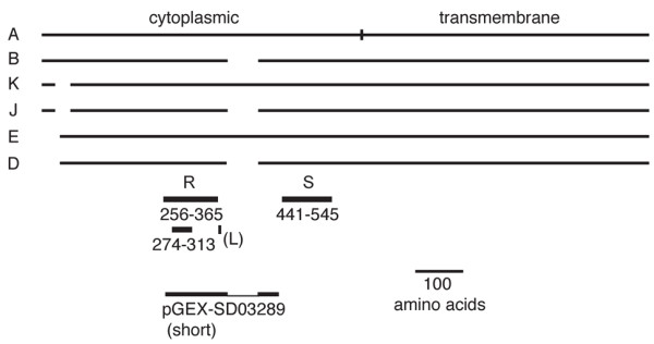
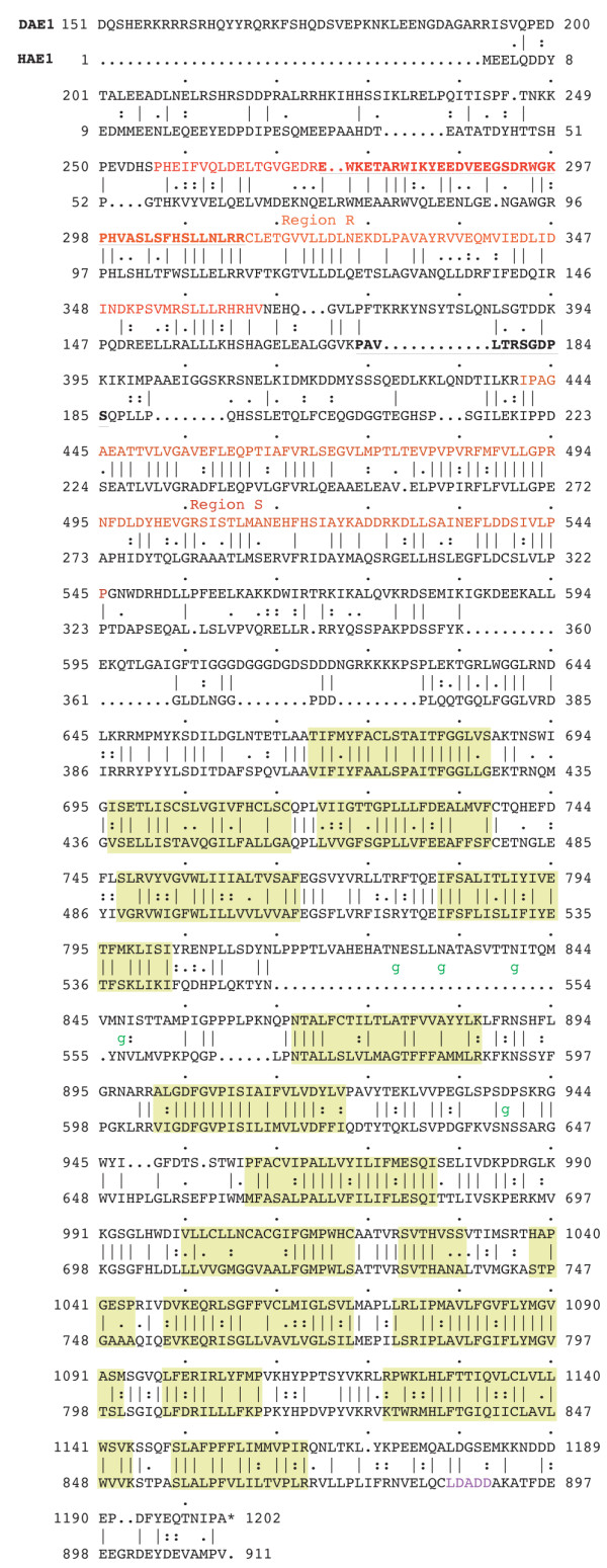
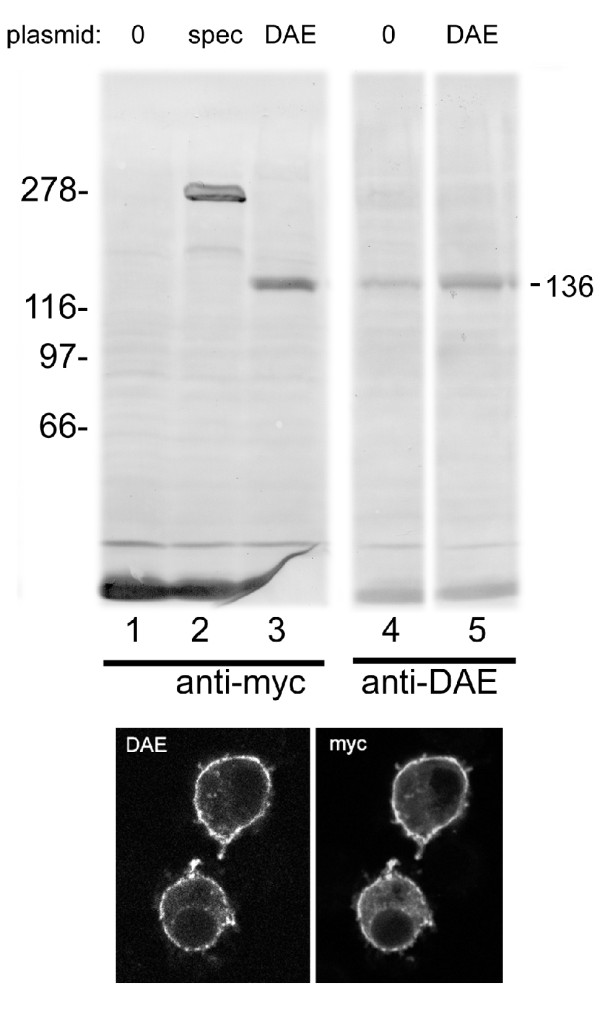
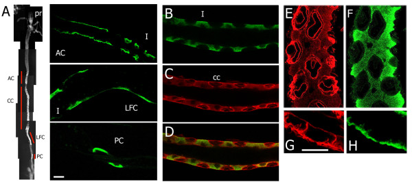
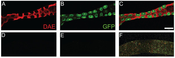
Results: Sequence comparisons established that DAE belongs to the SLC4A1-3 subfamily of anion exchangers that includes human AE1. Striking sequence conservation was observed in the C-terminal membrane transport domain and parts of the N-terminal cytoplasmic domain, but not in the proposed ankyrin-binding site. Using an antibody raised against DAE and a recombinant transgene expressed in Drosophila S2 cells DAE was shown to be a 136 kd plasma membrane protein. A major site of expression was found in the stomach acid-secreting region of the larval midgut. DAE codistributed with an infolded subcompartment of the basal plasma membrane of interstitial cells. However, spectrin did not codistribute with DAE at this site or in anterior midgut cells that abundantly expressed both spectrin and DAE. Ubiquitous knockdown of DAE with dsRNA eliminated antibody staining and was lethal, indicating that DAE is an essential gene product in Drosophila.

Conclusions: Based on the lack of colocalization and the lack of sequence conservation at the ankyrin-binding site, it appears that the well-characterized interaction between AE1 and the spectrin cytoskeleton in erythrocytes is not conserved in Drosophila. The results establish a pattern in which most of the known interactions between the spectrin cytoskeleton and the plasma membrane in mammals do not appear to be conserved in Drosophila.


Figures

Figure 1
Figure 1
Alternate protein products of the Drosophila anion exchanger gene. The six major classes of DAE protein products are depicted relative to the longest class (A). Classes K and J use an alternate splice site that removes part of the coding sequence near the N-terminus. Classes E and D use an alternate transcription start site and an internal methionine start codon. Classes B, J and D splice out an alternate exon which is located between two zones with high sequence identity (R and S). The short loop sequence (L) which is responsible for the interaction between AE1 and ankyrin in humans is not conserved in DAE. The boundaries of a short recombinant fragment of DAE expressed as a pGEX fusion protein are also shown.
Figure 2
Figure 2
Amino acid sequence alignment between DAE and human erythrocyte AE1. The positions of 16 predicted transmembrane sequences are indicated in green boxes. The boundaries of the conserved cytoplasmic domain sequences R and S are indicated in red. The conserved subregion R' is indicated in bold red type. The sequence of the ankyrin-binding site in human AE1 is indicated in bold black type. Consensus glycosylation sites in the linker between transmembrane regions [5,6] and [7,8] are each marked by g. The site of interaction between the C-terminal domain of AE1 and carbonic anhydrase is indicated in purple.
Figure 3
Figure 3
Expression of endogenous and recombinant anion exchanger in S2 tissue culture cells. Western blots of total S2 cell proteins were stained with mouse anti-myc epitope antibody (lanes 1-3) or affinity pure rabbit anti-DAE antibody (lanes 4-5). Untransfected cells (0) were compared to transfected cells expressing myc-tagged recombinant β spectrin as a control (lane 2) or myc-tagged recombinant DAE (lanes 3 & 5). The predicted size of the anion exchanger was 136 kD. Transfected S2 cells expressing myc-tagged DAE were stained with the same antibodies and fluorescent secondary antibodies (bottom panels). Staining was primarily observed at the plasma membrane including filopodia.
Figure 4
Figure 4
Immunolocalization of DAE in the larval midgut. A) Four distinct domains of DAE antibody-labeled cells in larval midgut: anterior cells (AC) upstream of the copper cell domain (CC), large flat cells (LFC) immediately downstream of the CC domain, and 2-3 posterior cells (PC) downstream of the LFC (Pr = proventriculus). The higher magnification views (bar = 10 um) show continuous labeling of the basal membrane of adjacent cells in the AC region (vs. interstitial cells (I) that alternate with copper cells downstream). LFCs were usually visible on only one side of the epithelial tube, downstream of the last interstitial cell. B) DAE staining in the copper cell region revealed a pattern of labeled interstitial cells (I) separated by unlabeled copper cells. C) The copper cells stained with anti-α spectrin antibody appeared as lozenge shapes with relatively bright staining of the basolateral region (merged in D). Higher magnification views of the α spectrin (E) and DAE (F) staining patterns emphasize their lack of overlap. Alpha spectrin was most conspicuous in the basolateral zone of contact between copper cells and interstitial cells and in the banana-shaped apical invagination of copper cells. In contrast, DAE was most conspicuous within the basal cytoplasm of interstitial cells and extended apically in a gradient. G&H) High magnification views of the anterior cells stained for α spectrin (G) or DAE (H) reveals their overlapping distribution in the basal membrane region, but not in the lateral region of cell-cell contact (Bar = 10 um).
Figure 5
Figure 5
RNAi knockdown of DAE expression. The specificity of the DAE antibody was tested by knocking down its expression with RNAi. The midgut-specific Mex-Gal4 driver was used to induce RNAi. In the cross scheme used RNAi-expressing larvae were distinguished from non-expressing siblings by the presence of a GFP reporter in the latter. Larvae were sorted by GFP expression and then dissected and stained with the anti-DAE antibody followed by Texas Red labeled secondary antibody. Control larvae carrying the GFP-marked balancer chromosome exhibited the expected pattern of interstitial cell DAE staining (A) with the GFP reporter fortuitously expressed in neighboring copper cells (B). Siblings that expressed UAS-RNAi (recognized by lack of GFP; E) showed no detectable DAE staining (D), indicating that the antibody was specific for DAE. The merged image (F) was overexposed to demonstrate the presence of the middle midgut. Bar = 20 um.

Similar articles

Cited by

References

    1. Lux SE, Palek J. In: Blood: Principles and practice of hematology. Handin RI, Lux SE, Stossel TP, editor. Philadelphia: J.B. Lippincott Co.; 1995. Disorders of the Red Cell Membrane; pp. 1701–1818.
    1. Bennett V, Baines AJ. Spectrin and ankyrin-based pathways: Metazoan inventions for integrating cells into tissues. Physiol Rev. 2001. pp. 1353–1388. - PubMed
    1. Dubreuil RR. Functional links between membrane transport and the spectrin cytoskeleton. J Membrane Biology. 2006;211:151–161. doi: 10.1007/s00232-006-0863-y. - DOI - PubMed
    1. Bennett V, Healy J. Organizing the fluid membrane bilayer: diseases linked to spectrin and ankyrin. Trends in Molecular Medicine. 2008;14:28–36. doi: 10.1016/j.molmed.2007.11.005. - DOI - PubMed
    1. Dubreuil RR. In: Advance in Molecular Cell Biology. Khurana S, editor. Vol. 37. New York:Elsevier; 2006. Spectrin function: A survey of genetic systems from Drosophila to humans; pp. 68–88.

Publication types

MeSH terms

LinkOut - more resources