. 2010 Jun 23:7:55.
doi: 10.1186/1742-4690-7-55.

Semen-mediated enhancement of HIV infection is donor-dependent and correlates with the levels of SEVI

Affiliations

Semen-mediated enhancement of HIV infection is donor-dependent and correlates with the levels of SEVI

Kyeong-Ae Kim et al. Retrovirology. .

Abstract

Background: HIV-1 is usually transmitted in the presence of semen. We have shown that semen boosts HIV-1 infection and contains fragments of prostatic acid phosphatase (PAP) forming amyloid aggregates termed SEVI (semen-derived enhancer of viral infection) that promote virion attachment to target cells. Despite its importance for the global spread of HIV-1, however, the effect of semen on virus infection is controversial.

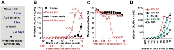
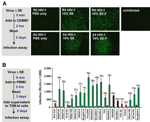
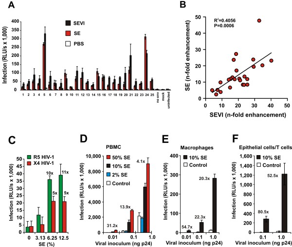
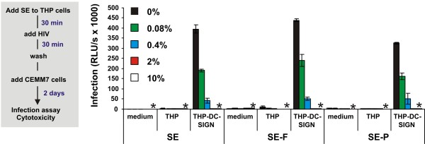
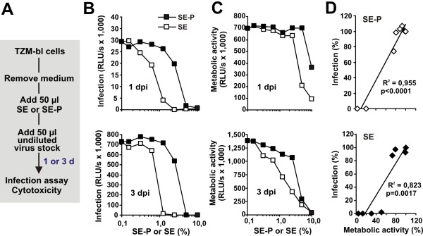
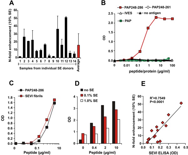
Results: Here, we established methods allowing the meaningful analysis of semen by minimizing its cytotoxic effects and partly recapitulating the conditions encountered during sexual HIV-1 transmission. We show that semen rapidly and effectively enhances the infectivity of HIV-1, HIV-2, and SIV. This enhancement occurs independently of the viral genotype and coreceptor tropism as well as the virus producer and target cell type. Semen-mediated enhancement of HIV-1 infection was also observed under acidic pH conditions and in the presence of vaginal fluid. We further show that the potency of semen in boosting HIV-1 infection is donor dependent and correlates with the levels of SEVI.

Conclusions: Our results show that semen strongly enhances the infectivity of HIV-1 and other primate lentiviruses and that SEVI contributes to this effect. Thus, SEVI may play an important role in the sexual transmission of HIV-1 and addition of SEVI inhibitors to microbicides may improve their efficacy.


Figures

Figure 1
Figure 1
Effect of SE on HIV infection. (A) Schema describing the experimental procedure. (B) Effect of treatment of virus stocks with 90% (v/v) of SE on R5 HIV-1 infection. TZM-bl cells were infected with the indicated dilutions of SE- or PBS-treated virus stocks. The inoculum was either removed after 2 hours of exposure (wash) or left on the cells. Shown are average β-galactosidase activities (n = 3) measured 2 days after virus exposure. RLU/s: relative light units per second. The numbers above the upper curve give n-fold enhancement of HIV infection by SE relative to that measured for the corresponding PBS control. (C) Metabolic activities of cells analysed in B. (D) Effect of low concentrations of SE on HIV infection. R5 HIV-1 stocks were treated with the indicated concentrations of SE, diluted and used to infect TZM-bl cells. The Y-axis gives average values of triplicate infections, and the X-axis gives the final dilution of the virus stocks. The infection levels were determined as described above. Percentages refer to the SE concentrations during virion treatment. The final concentrations in the cell culture are 15-fold lower.
Figure 2
Figure 2
Effect of exposure times and pH values on SE mediated enhancement of HIV infection. (A) Effect of pre-incubation time on SE-mediated HIV infection enhancement. R5 HIV-1 was mixed with the indicated concentrations of SE, incubated for the various time periods, and 20 μl of the virus stocks was used to infect 280 μl TZM-bl cell cultures in triplicates. Values in all panels give averages ± SD (n = 3) measured 3 days after virus exposure. (B) The SE/virus mixture was incubated for 10 min at RT, and 20 μl were added to 280 μl TZM-bl cells. After different time points, the supernatant was removed, and fresh DMEM was added for further culture. The star indicates high cytotoxicity. (C) Virus stocks of R5 HIV-1 treated with the indicated concentrations of SE were used to infect TZM-bl cultures adjusted to the indicated pH values. After two hours of virus exposure, the virus stocks were removed, and the cells were cultured in fresh medium under neutral pH conditions. Higher or lower pH values could not be analyzed as they were cytotoxic. Both panels give average values ± SD (n = 3). (D) Virus stocks adjusted to the indicated pH values were either treated with PBS or with various concentrations of SE and subsequently used to infect TZM-bl indicator cells. (E) TZM-bl cells were incubated with either PBS or 10% cervico vaginal lavage (CLF) and infected with medium or 10% SE treated HIV-1. Infection rates were determined 3 dpi.
Figure 3
Figure 3
The enhancing effect of SE is independent of the viral coreceptor tropism. (A) Analysis of CEMx-M7 cells infected with untreated or SE-treated (10% v/v) X4 and R5 HIV-1 by fluorescence microscopy 2 dpi. (B) Treatment with SE enhances HIV-1 infection of primary PBMCs. Stimulated PBMCs were infected with the same dose of the indicated HIV-1 NL4-3 V3 recombinants that were either not treated or treated with 10% (v/v) SE. Three days later, 100 μl of the cell-free PBMC culture supernatant was used to infect TZM-bl indicator cells. Shown are average infection rates of TZM-bl cell ± SD (n = 3) measured 2 days after virus exposure. X4 virus is color-coded red; R5 virus, green; and X4R5 HIV-1, black.
Figure 4
Figure 4
SE enhances founder HIV infection and boosts HIV infection independently of the viral producer and target cell type. (A) Effect of SE on HIV particles carrying gp120/41 from founder viruses. Pseudotyped HIV-1 particles were generated by transient transfection of 293T cells with an env-defective HIV-1 NL4-3 backbone and plasmids expressing the Env proteins previously described (34). Virions were treated with medium, 10 μg/ml SEVI or 10% SE and used to infect TZM-bl cells. The inoculum was removed after 2 hrs and infection rates were determined 2 dpi. Shown are the average levels of triplicate TZM-bl cell infections ± SD. (B) Correlation between the magnitudes of SEVI and SE-mediated enhancement of HIV-1 pseudotype infection shown in Fig 4A. N-fold enhanced infection rates were calculated by dividing infection rates obtained in the presence of SEVI or SE by those of mock-treated virus infection. (C) SE enhances infection of testis derived HIV-1. X4 tropic HIV-1 IIIb and R5 tropic SF162 were harvested from infected testis tissue, treated with indicated concentrations of SE and used to infect TZM-bl cells. (D, E) SE enhances the infectiousness of HIV-1 for PBMCs and macrophages. Stocks of an R5-tropic HIV-1 NL4-3 V3 variant (92TH04.12) containing the luciferase reporter gene in place of nef were generated by transient transfection of 293T cells. Virus stocks were treated with the indicated concentrations of SE and used to infect PBMC (D) and macrophages (E). Similar results were obtained using various primary HIV-1 strains. (F) SE favours in trans-infection of T cells by viral particles bound to epithelial cells. CaSki cells derived from an epithelial cervical carcinoma were exposed to HIV-1 treated with SE or medium for 3 hrs. Subsequently, the virus inoculum was washed out and the cells were cocultivated with CEM-M7 cells for three days. Infection rates were determined by luciferase assay. The numbers above the bars indicate n-fold enhancement relative to the infectivity measured using PBS/medium-treated virus stocks.
Figure 5
Figure 5
Semen inhibits trans-infection of T cells by DC-SIGN. B-THP-1-DC-SIGN cells were treated with the indicated concentration of SE, SE-F or SE-P for 30 min, subsequently exposed to R5 HIV-1 for 30 min, washed and cocultivated with CEM-M7 cells. The levels of infection mediated by B-THP-1 cells, which do not express DC-SIGN, and the absence of cells (medium) are also shown as controls. Shown are average values ± SD derived from triplicate infections. Stars indicate cytotoxicity, infection rates obtained after infection with 2% and 10% SE, SE-F or SE-P treated virus were close to background luciferase activities of uninfected cells.
Figure 6
Figure 6
Adding SE or SE-P directly to target cells results in reduced metabolic activity and HIV infection rates. (A) Schematic outline of the experiment. TZM-bl cells were incubated with different dilutions of SE or SE-P and subsequently infected with HIV-1. (B) Infection rates and (C) metabolic activities were determined after 1 (upper panel) or 3 days (lower panel). (D) Correlation between Tat-driven reporter activities ("infection") and the metabolic activities of the target cells. Values were derived from the experiments shown in panels B and C, and are shown relative to those obtained in the absence of SE or SE-P (100%).
Figure 7
Figure 7
The HIV enhancing activity of individual SE samples correlates with SEVI concentrations. (A) Enhancement of HIV-1 infection by SE samples from 14 different donors. R5 HIV-1 stocks were mixed with 10% (v/v) of the SE samples or PBS and used for infection of TZM-bl indicator cells. Similar results were obtained using SE samples from more than 80 additional donors. Reactivity of anti-SEVI-antiserum from guinea pigs to (B) the indicated monomeric peptides or IVES, a peptide containing the reverse amino acid sequence of PAP248-286 and full-length PAP. (C) SEVI fibrils or (D) pooled SE spiked with SEVI. Similar results were obtained using an antiserum from rabbits. (E) Correlation between the magnitude of enhancement of HIV-1 infection by individual SE samples and the quantity of SEVI/PAP248-286 detectable using the anti-SEVI antiserum.

References

    1. Korber B, Muldoon M, Theiler J, Gao F, Gupta R, Lapedes A, Hahn BH, Wolinsky S, Bhattacharya T. Timing the ancestor of the HIV-1 pandemic strains. Science. 2000;288:1789–96. doi: 10.1126/science.288.5472.1789. - DOI - PubMed
    1. UNAIDS. 2008 AIDS epidemic update. http://www.unaids.org
    1. Gray RH, Wawer MJ, Brookmeyer R, Sewankambo NK, Serwadda D, Wabwire-Mangen F, Lutalo T, Li X, vanCott T, Quinn TC. Rakai Project Team. Probability of HIV-1 transmission per coital act in monogamous, heterosexual, HIV-1-discordant couples in Rakai, Uganda. Lancet. 2001;357:1149–53. doi: 10.1016/S0140-6736(00)04331-2. - DOI - PubMed
    1. Padian NS, Shiboski SC, Glass SO, Vittinghoff E. Heterosexual transmission of human immunodeficiency virus (HIV) in northern California: results from a ten-year study. Am J Epidemiol. 1997;146:350–57. - PubMed
    1. Vittinghoff E, Douglas J, Judson F, McKirnan D, MacQueen K, Buchbinder S. Per-contact risk of human immunodeficiency virus transmission between male sexual partners. Am J Epidemiol. 1999;150:306–11. - PubMed

Publication types